1NYK image
Deposition Date 2003-02-12
Release Date 2003-06-24
Last Version Date 2024-11-20
Entry Detail
PDB ID:
1NYK
Title:
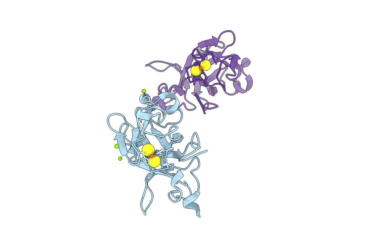
Crystal Structure of the Rieske protein from Thermus thermophilus
Biological Source:
Source Organism:
Host Organism:
Method Details:
Experimental Method:
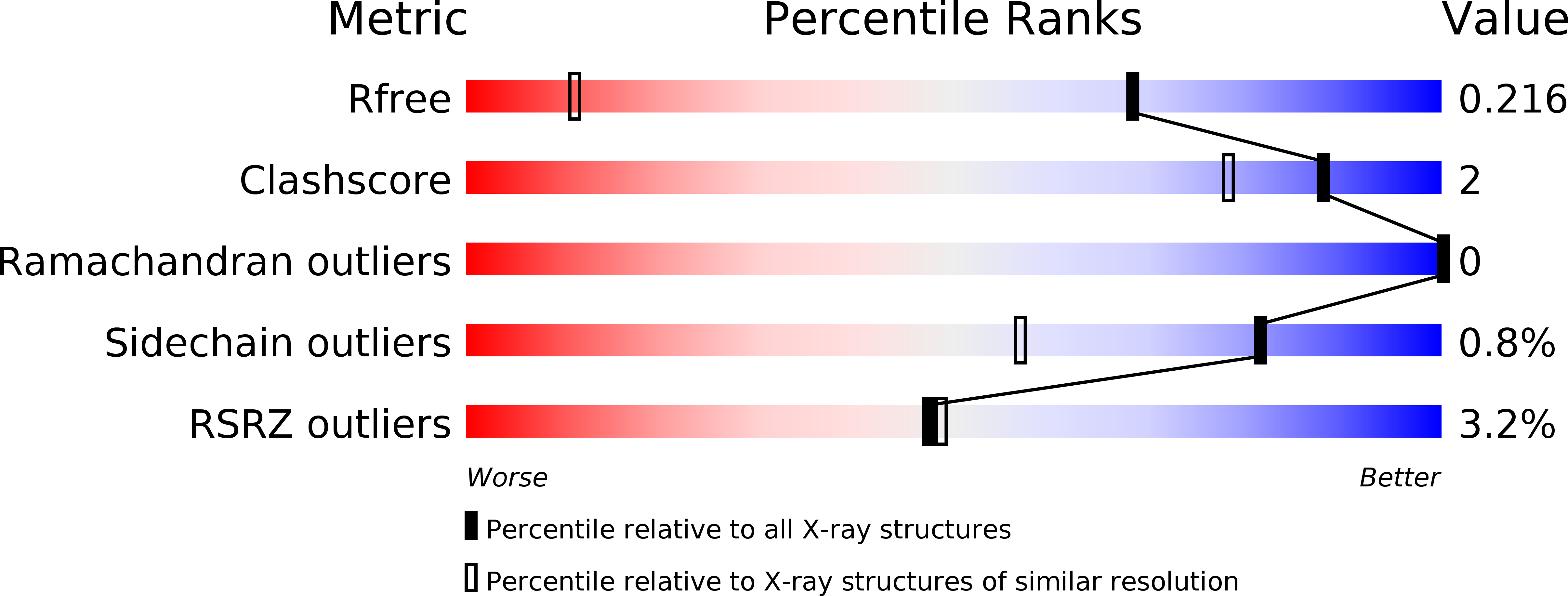
Resolution:
1.31 Å
R-Value Free:
0.21
R-Value Work:
0.15
R-Value Observed:
0.15
Space Group:
C 2 2 21
Macromolecular Entities
Structures with similar UniProt ID
Protein Blast
Polymer Type:polypeptide(L)
Molecule:Rieske iron-sulfur protein
Chain IDs:A, B
Chain Length:165
Number of Molecules:2
Biological Source:Thermus thermophilus
Primary Citation
High resolution structure of the soluble, respiratory-type Rieske protein from Thermus thermophilus: Analysis and Comparison
Biochemistry 42 7303 7317 (2003)
PMID: 12809486 DOI: 10.1021/bi0342719

Abstact

The structure of the soluble Rieske protein from Thermus thermophilus has been determined at a resolution of 1.3 A at pH 8.5 using multiwavelength anomalous dispersion (MAD) techniques. This is the first report of a Rieske protein from a menaquinone-utilizing organism. The structure shows an overall fold similar to previously reported Rieske proteins. A novel feature of this crystal form appears to be a shared hydrogen between the His-134 imidazole ring ligated to Fe2 of the [2Fe-2S] cluster and its symmetry partner, His-134', one being formally an imidazolate anion, Fe2-(His-134)N(epsilon)(-)...H-N(epsilon')(His-134')-Fe2', in which crystallographic C(2) axes pass equidistant between N(epsilon)...N(epsilon') and normal to the line defined by N(epsilon)...N(epsilon'). This provides evidence for a stable, oxidized cluster with a His(-) ligand and lends support to a previously proposed mechanism of coupled proton and electron transfer. A detailed comparison of the Thermus Rieske protein with six other Rieske and Rieske-type proteins indicates: (a) The cluster binding domain is tightly conserved. (b) The 3-D structure of the 10 beta-strand fold is conserved, even among the most divergent proteins. (c) There is an approximately linear relation between acid-pH redox potential and number of H-bonds to the cluster. (d) These proteins have two faces, one points into the larger complex (bc(1), b(6)f, or other), is involved in the proton coupled electron transfer function, and is highly conserved. The second is oriented toward the solvent and shows wide variation in charge, sequence, length, hydrophobicity, and secondary elements in the loops that connect the beta-sheets.

Legend

Protein

Chemical

Disease

Primary Citation of related structures