Deposition Date
2003-02-12
Release Date
2003-06-24
Last Version Date
2024-02-14
Entry Detail
PDB ID:
1NYH
Keywords:
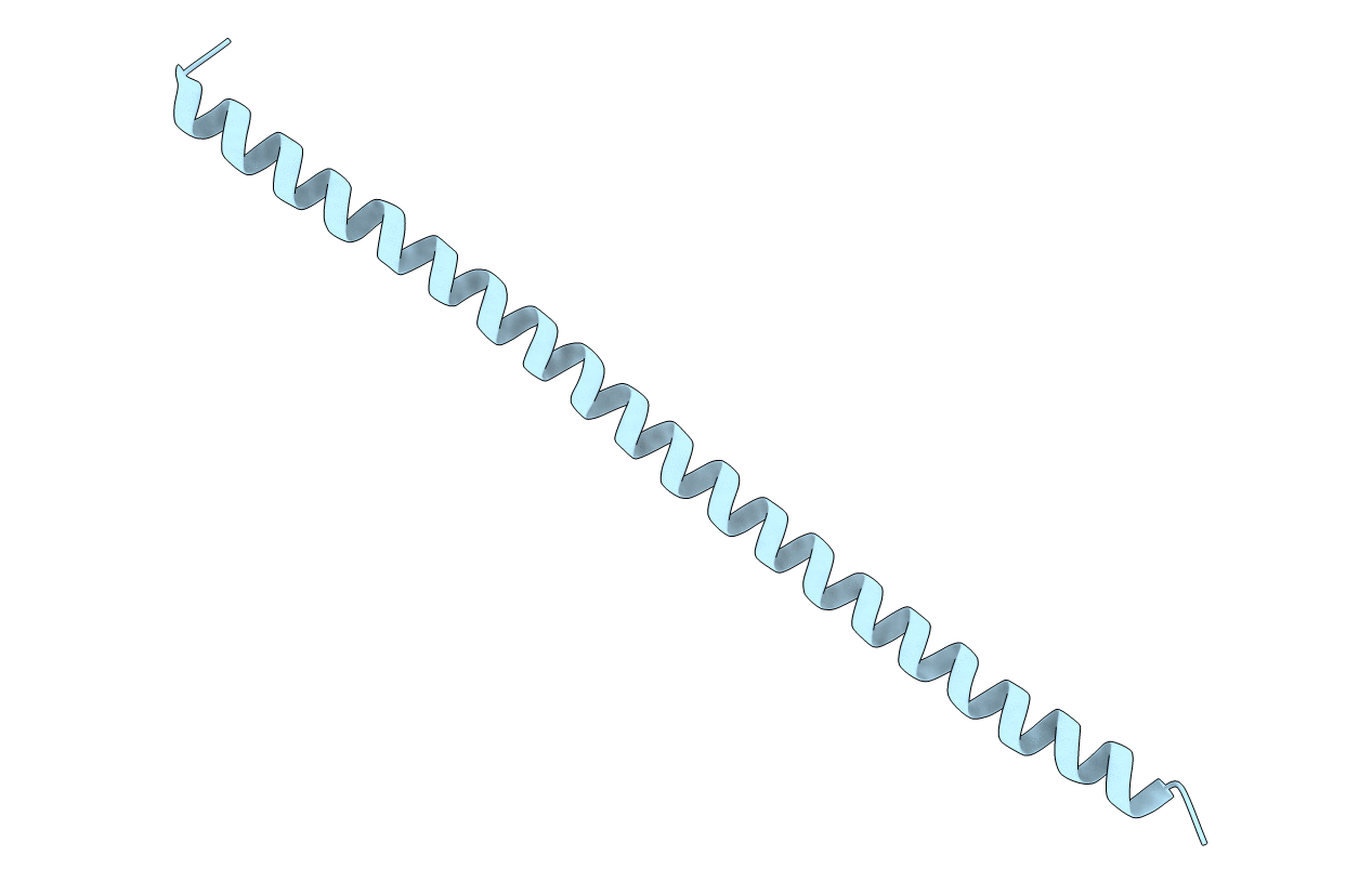
Title:
Crystal Structure of the Coiled-coil Dimerization Motif of Sir4
Biological Source:
Source Organism(s):
Saccharomyces cerevisiae (Taxon ID: 4932)
Expression System(s):
Method Details:
Experimental Method:
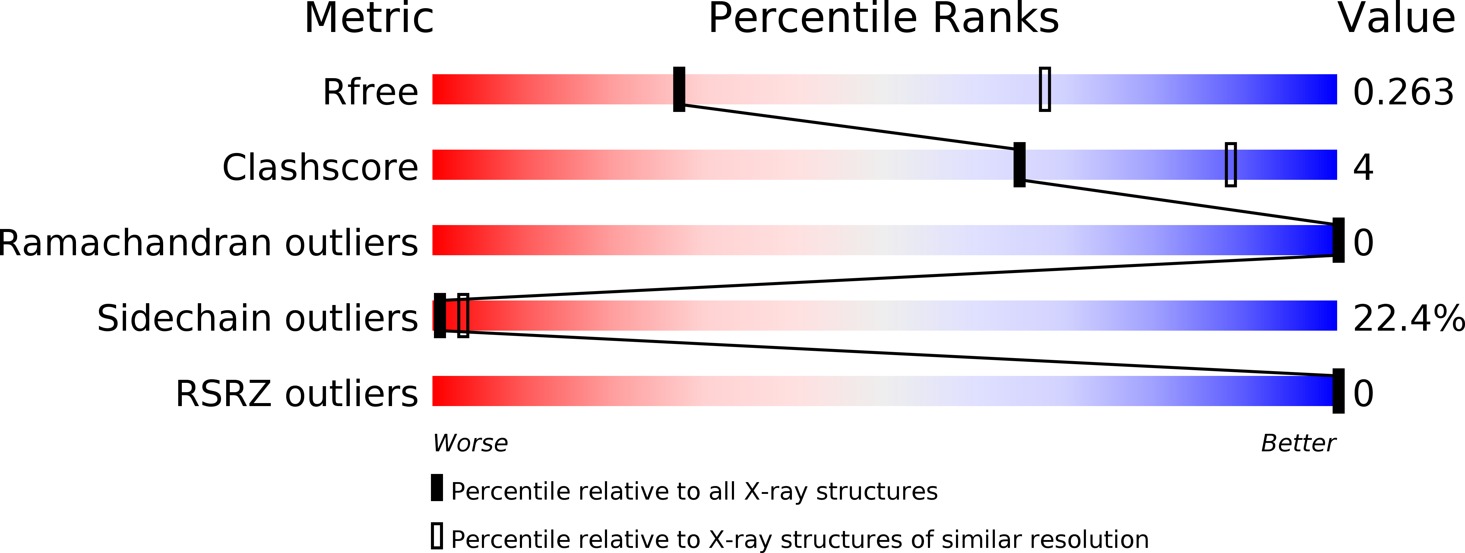
Resolution:
3.10 Å
R-Value Free:
0.28
R-Value Work:
0.27
R-Value Observed:
0.27
Space Group:
P 65 2 2