Deposition Date
2003-02-10
Release Date
2004-06-15
Last Version Date
2024-05-29
Entry Detail
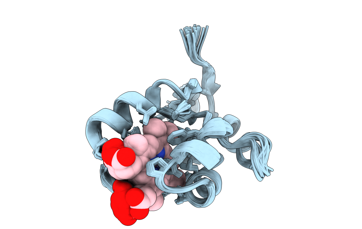
PDB ID:
1NX7
Keywords:
Title:
Solution Structure of Oxidized Bovine Microsomal Cytochrome B5
Biological Source:
Source Organism(s):
Bos taurus (Taxon ID: 9913)
Expression System(s):
Method Details:
Experimental Method:
Conformers Submitted:
30