1NWZ image
Deposition Date 2003-02-07
Release Date 2003-03-11
Last Version Date 2025-03-26
Entry Detail
PDB ID:
1NWZ
Title:
PYP Ultra-high resolution structure of a Bacterial Photoreceptor
Biological Source:
Source Organism:
Method Details:
Experimental Method:
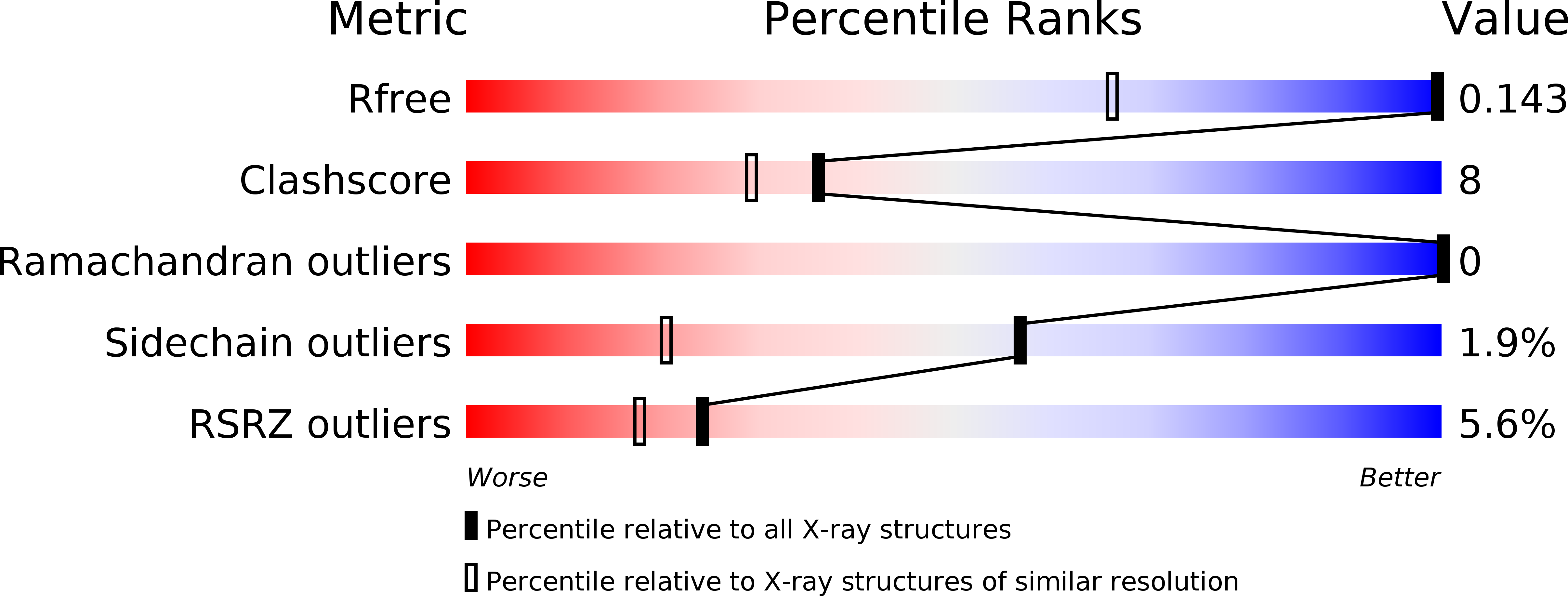
Resolution:
0.82 Å
R-Value Free:
0.14
R-Value Work:
0.12
R-Value Observed:
0.12
Space Group:
P 63
Macromolecular Entities
Structures with similar UniProt ID
Protein Blast
Polymer Type:polypeptide(L)
Molecule:Photoactive yellow protein
Gene (Uniprot):pyp
Chain IDs:A
Chain Length:125
Number of Molecules:1
Biological Source:Halorhodospira halophila
Ligand Molecules
Primary Citation
Anticipatory active-site motions and chromophore distortions prime photoreceptor PYP for light activation
Nat.Struct.Biol. 10 663 668 (2003)
PMID: 12872160 DOI: 10.1038/nsb958

Abstact

Protein photoreceptors use small-molecule cofactors called chromophores to detect light. Only under the influence of the receptors' active sites do these chromophores adopt spectral and photochemical properties that suit the receptors' functional requirements. This protein-induced change in chromophore properties is called photochemical tuning and is a prime example for the general--but poorly understood--process of chemical tuning through which proteins shape the reactivity of their active-site groups. Here we report the 0.82-A resolution X-ray structure of the bacterial light receptor photoactive yellow protein (PYP). The unusually precise structure reveals deviations from expected molecular geometries and anisotropic atomic displacements in the PYP active site. Our analysis of these deviations points directly to the intramolecular forces and active-site dynamics that tune the properties of PYP's chromophore to absorb blue light, suppress fluorescence, and favor the required light-driven double-bond isomerization.

Legend

Protein

Chemical

Disease

Primary Citation of related structures