
Deposition Date
2003-02-06
Release Date
2003-04-08
Last Version Date
2024-05-22
Entry Detail
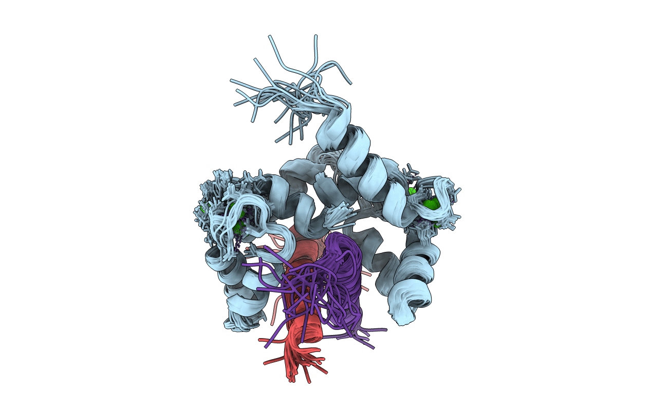
PDB ID:
1NWD
Keywords:
Title:
Solution Structure of Ca2+/Calmodulin bound to the C-terminal Domain of Petunia Glutamate Decarboxylase
Biological Source:
Source Organism(s):
Xenopus laevis (Taxon ID: 8355)
Petunia x hybrida (Taxon ID: 4102)
Petunia x hybrida (Taxon ID: 4102)
Expression System(s):
Method Details:
Experimental Method:
Conformers Calculated:
200
Conformers Submitted:
20
Selection Criteria:
structures with the lowest energy
