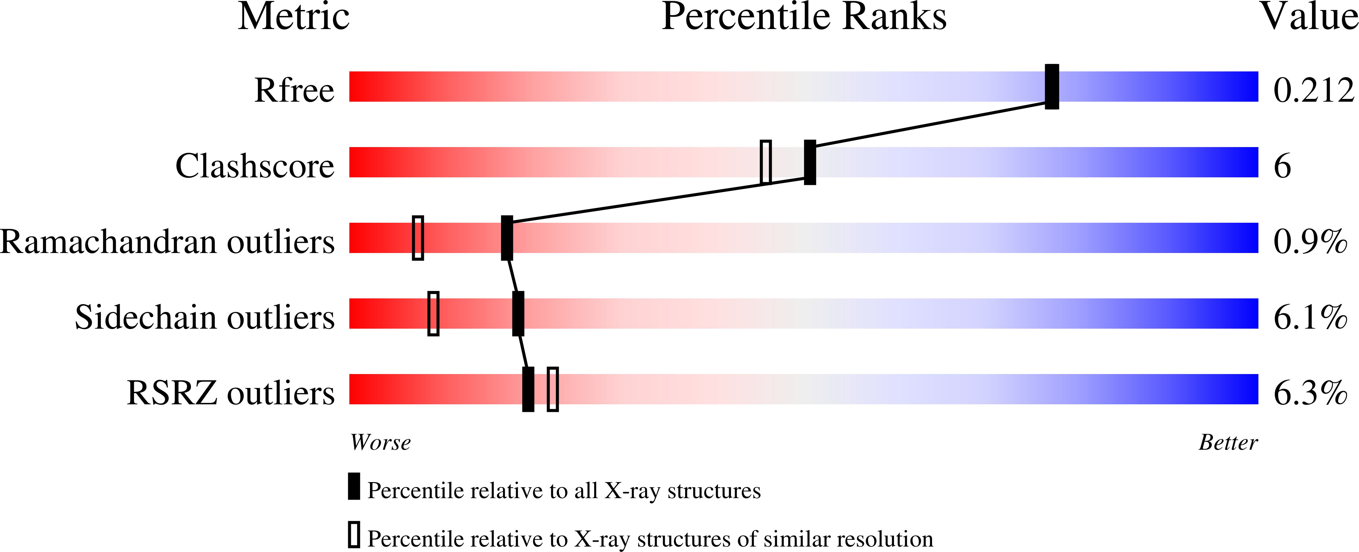
1NVI image
Deposition Date 2003-02-03
Release Date 2003-05-06
Last Version Date 2023-08-16
Entry Detail
PDB ID:
1NVI
Keywords:
Title:
Orthorhombic Crystal Form of Molybdopterin Synthase
Biological Source:
Source Organism:
Escherichia coli (Taxon ID: 562)
Method Details:
Experimental Method:
Resolution:
1.90 Å
R-Value Free:
0.20
R-Value Work:
0.17
R-Value Observed:
0.17
Space Group:
I 21 21 21
Macromolecular Entities
Structures with similar UniProt ID
Protein Blast
Polymer Type:polypeptide(L)
Molecule:Molybdopterin converting factor subunit 1
Gene (Uniprot):moaD
Mutagens:I2V
Chain IDs:A (auth: D)
Chain Length:81
Number of Molecules:1
Biological Source:Escherichia coli
Structures with similar UniProt ID
Protein Blast
Polymer Type:polypeptide(L)
Molecule:Molybdopterin converting factor subunit 2
Gene (Uniprot):moaE
Chain IDs:B (auth: E)
Chain Length:150
Number of Molecules:1
Biological Source:Escherichia coli
Primary Citation
Structural Studies of Molybdopterin Synthase Provide Insights into its Catalytic Mechanism
J.Biol.Chem. 278 14514 14522 (2003)
PMID: 12571227 DOI: 10.1074/jbc.M300449200

Abstact

Molybdenum cofactor biosynthesis is an evolutionarily conserved pathway present in eubacteria, archaea, and eukaryotes, including humans. Genetic deficiencies of enzymes involved in cofactor biosynthesis in humans lead to a severe and usually fatal disease. The molybdenum cofactor contains a tricyclic pyranopterin, termed molybdopterin, that bears the cis-dithiolene group responsible for molybdenum ligation. The dithiolene group of molybdopterin is generated by molybdopterin synthase, which consists of a large (MoaE) and small (MoaD) subunit. The crystal structure of molybdopterin synthase revealed a heterotetrameric enzyme in which the C terminus of each MoaD subunit is deeply inserted into a MoaE subunit to form the active site. In the activated form of the enzyme, the MoaD C terminus is present as a thiocarboxylate. The present study identified the position of the thiocarboxylate sulfur by exploiting the anomalous signal originating from the sulfur atom. The structure of molybdopterin synthase in a novel crystal form revealed a binding pocket for the terminal phosphate of molybdopterin, the product of the enzyme, and suggested a binding site for the pterin moiety present in precursor Z and molybdopterin. Finally, the crystal structure of the MoaE homodimer provides insights into the conformational changes accompanying binding of the MoaD subunit.

Legend

Protein

Chemical

Disease

Primary Citation of related structures