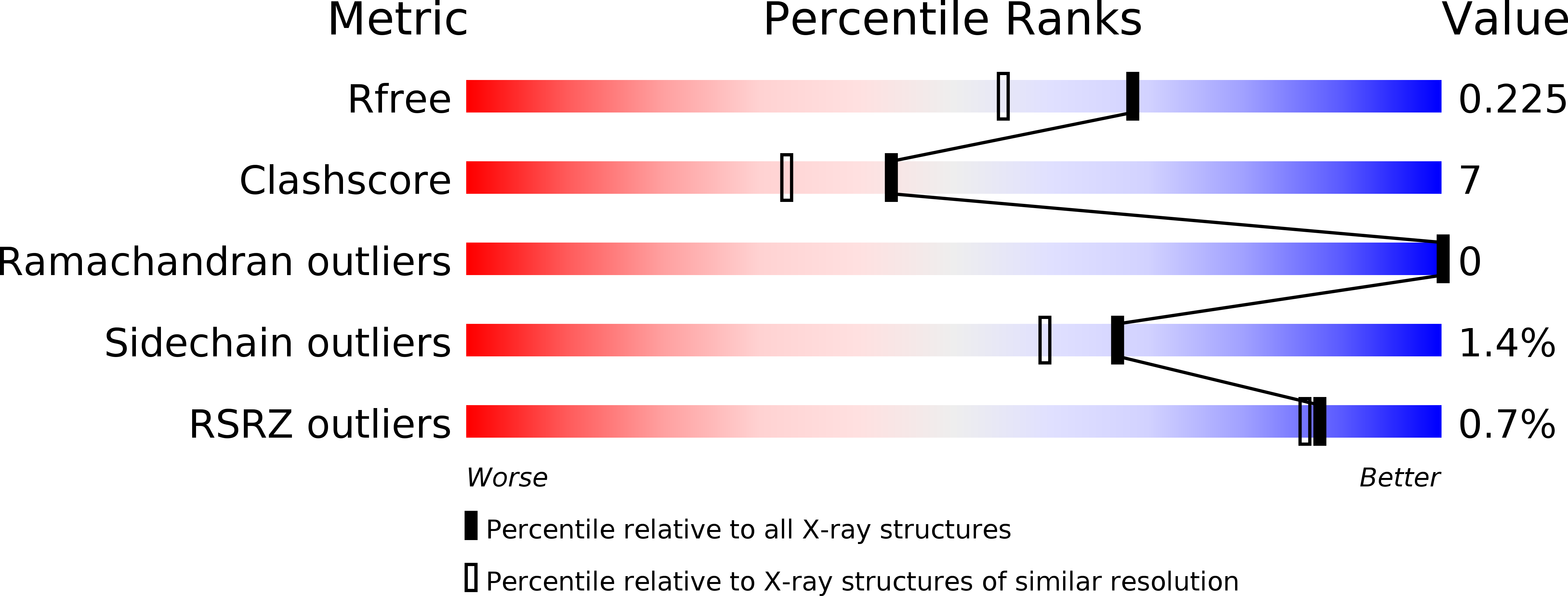
1NUL image
Deposition Date 1996-10-15
Release Date 1997-05-15
Last Version Date 2024-02-14
Entry Detail
PDB ID:
1NUL
Title:
XPRTASE FROM E. COLI
Biological Source:
Source Organism:
Escherichia coli (Taxon ID: 562)
Method Details:
Experimental Method:
Resolution:
1.80 Å
R-Value Free:
0.23
R-Value Work:
0.19
R-Value Observed:
0.19
Space Group:
P 21 21 2
Macromolecular Entities
Structures with similar UniProt ID
Protein Blast
Polymer Type:polypeptide(L)
Molecule:XANTHINE-GUANINE PHOSPHORIBOSYLTRANSFERASE
Gene (Uniprot):gpt
Chain IDs:A, B
Chain Length:152
Number of Molecules:2
Biological Source:Escherichia coli
Primary Citation
Crystal structure of Escherichia coli xanthine phosphoribosyltransferase.
Biochemistry 36 4125 4134 (1997)
PMID: 9100006 DOI: 10.1021/bi962640d

Abstact

Xanthine phosphoribosyltransferase (XPRT; EC 2.4.2.22) from Escherichia coli is a tetrameric enzyme having 152 residues per subunit. XPRT catalyzes the transfer of the phosphoribosyl group from 5-phospho-alpha-D-ribosyl 1-pyrophosphate (PRib-PP) to the 6-oxopurine bases guanine, xanthine, and hypoxanthine to form GMP, XMP, and IMP, respectively. Crystals grown in the absence of substrate or product were used to determine the structure of XPRT at a resolution of 1.8 A, by multiple isomorphous replacement. The core structure of XPRT includes a five-stranded parallel beta-sheet surrounded by three alpha-helices, which is similar to that observed in other known phosphoribosyltransferase (PRTase) structures. The XPRT structure also has several interesting features. A glutamine residue in the purine binding site may be responsible for the altered 6-oxopurine base specificity seen in this enzyme compared to other 6-oxopurine PRTases. Also, we observe both a magnesium ion and a sulfate ion bound at the PRib-PP binding site of XPRT. The sulfate ion interacts with Arg-37 which has a cis-peptide conformation, and the magnesium ion interacts with Asp-89, a highly conserved acidic residue in the PRib-PP binding site motif. The XPRT structure also incorporates a feature which has not been observed in other PRTase structures. The C-terminal 12 residues of XPRT adopt an unusual extended conformation and make interactions with a neighboring subunit. The very last residue, Arg-152, could form part of the active site of a symmetry-related subunit in the XPRT tetramer.

Legend

Protein

Chemical

Disease

Primary Citation of related structures