Deposition Date
2003-01-30
Release Date
2003-10-07
Last Version Date
2024-11-06
Entry Detail
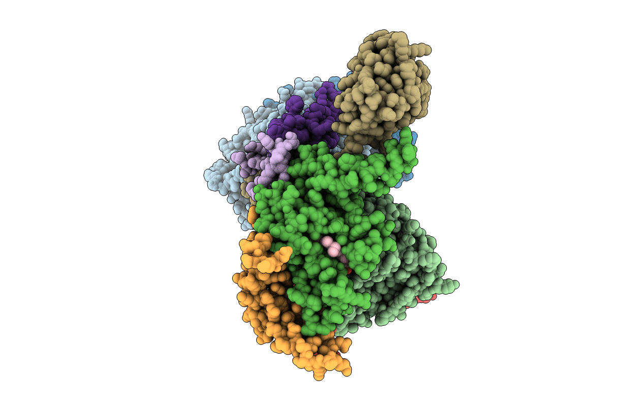
PDB ID:
1NTZ
Keywords:
Title:
Crystal Structure of Mitochondrial Cytochrome bc1 Complex Bound with Ubiquinone
Biological Source:
Source Organism(s):
Bos taurus (Taxon ID: 9913)
Method Details:
Experimental Method:
Resolution:
2.60 Å
R-Value Free:
0.28
R-Value Work:
0.24
R-Value Observed:
0.24
Space Group:
I 41 2 2