Deposition Date
1995-07-04
Release Date
1995-10-15
Last Version Date
2024-02-14
Entry Detail
PDB ID:
1NSK
Keywords:
Title:
THE CRYSTAL STRUCTURE OF A HUMAN NUCLEOSIDE DIPHOSPHATE KINASE, NM23-H2
Biological Source:
Source Organism(s):
Homo sapiens (Taxon ID: 9606)
Expression System(s):
Method Details:
Experimental Method:
Resolution:
2.80 Å
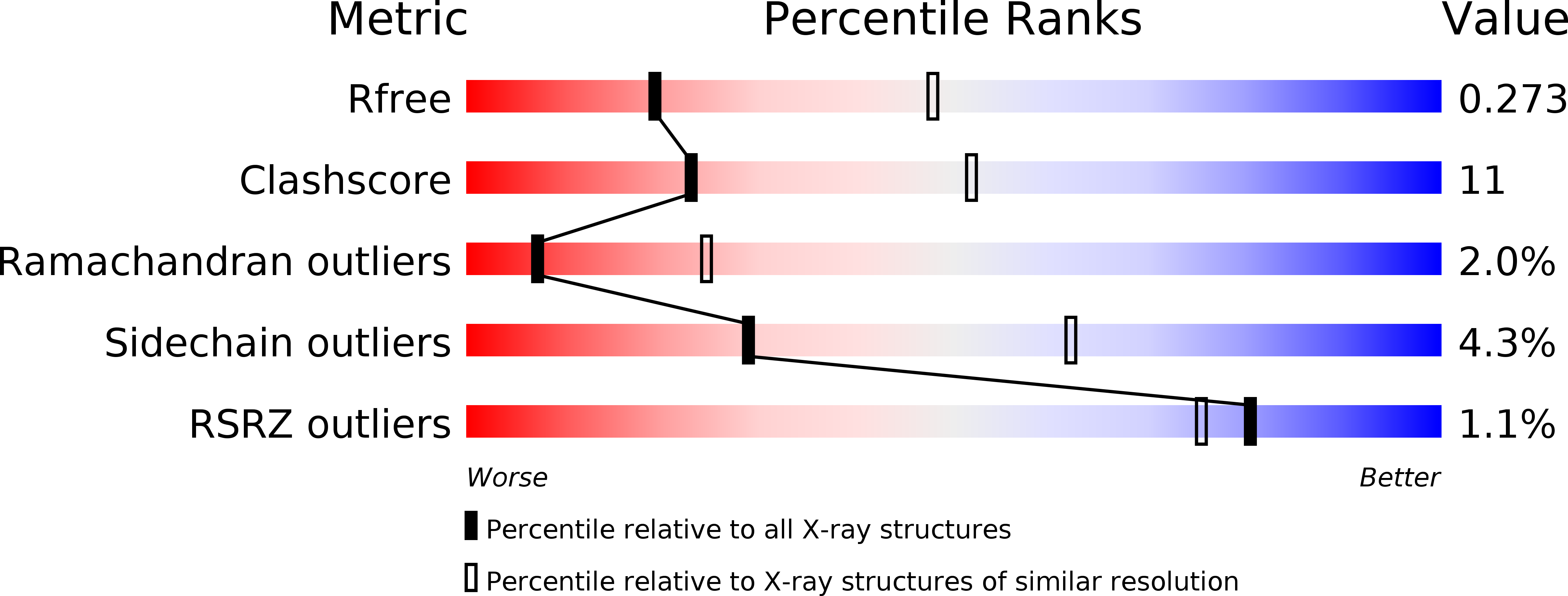
R-Value Free:
0.29
R-Value Work:
0.24
R-Value Observed:
0.24
Space Group:
P 21 21 21