1NSH image
Deposition Date 2003-01-27
Release Date 2003-07-15
Last Version Date 2024-05-22
Entry Detail
PDB ID:
1NSH
Title:
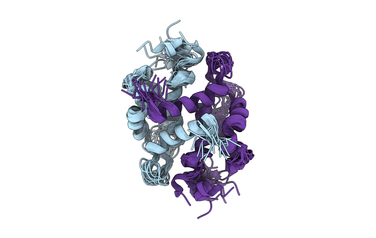
Solution Structure of Rabbit apo-S100A11 (19 models)
Biological Source:
Source Organism:
Method Details:
Experimental Method:
Conformers Calculated:
100
Conformers Submitted:
19
Selection Criteria:
structures with the lowest energy
Macromolecular Entities
Polymer Type:polypeptide(L)
Molecule:Calgizzarin
Gene (Uniprot):S100A11
Chain IDs:A, B
Chain Length:101
Number of Molecules:2
Biological Source:Oryctolagus cuniculus
Ligand Molecules
Primary Citation
Unmasking the Annexin I Interaction from the structure of apo-S100A11
Structure 11 887 897 (2003)
PMID: 12842051 DOI: 10.1016/S0969-2126(03)00126-6

Abstact

S100A11 is a homodimeric EF-hand calcium binding protein that undergoes a calcium-induced conformational change and interacts with the phospholipid binding protein annexin I to coordinate membrane association. In this work, the solution structure of apo-S100A11 has been determined by NMR spectroscopy to uncover the details of its calcium-induced structural change. Apo-S100A11 forms a tight globular structure having a near antiparallel orientation of helices III and IV in calcium binding site II. Further, helices I and IV, and I and I', form a more closed arrangement than observed in other apo-S100 proteins. This helix arrangement in apo-S100A11 partially buries residues in helices I (P3, E11, A15), III (V55, R58, M59), and IV (A86, C87, S90) and the linker (A45, F46), which are required for interaction with annexin I in the calcium-bound state. In apo-S100A11, this results in a "masked" binding surface that prevents annexin I binding but is uncovered upon calcium binding.

Legend

Protein

Chemical

Disease

Primary Citation of related structures