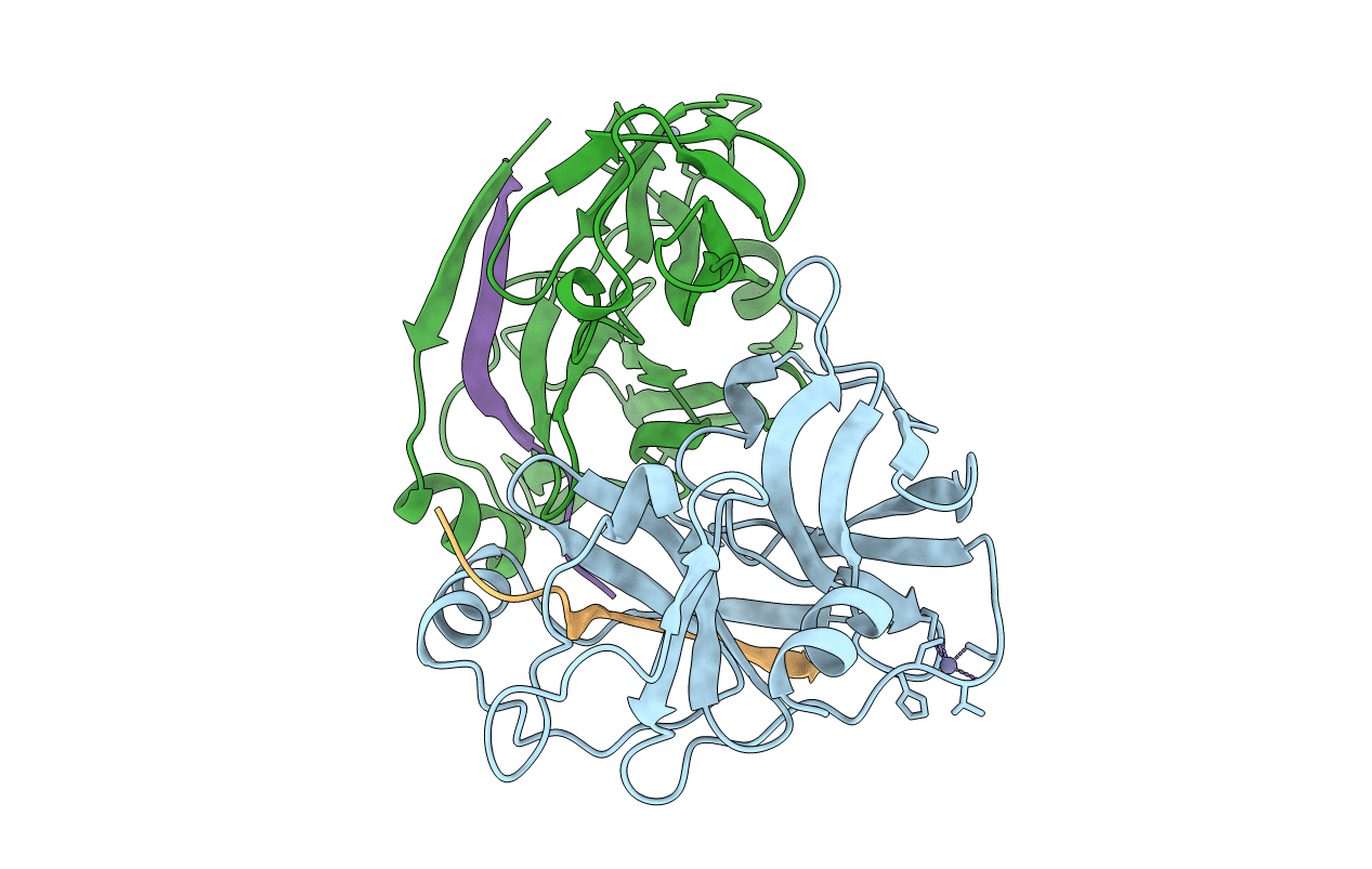
1NS3 image
Entry Detail
PDB ID:
1NS3
Title:
STRUCTURE OF HCV PROTEASE (BK STRAIN)
Biological Source:
Source Organism:
Host Organism:
PDB Version:
Deposition Date:
1997-04-05
Release Date:
1998-04-08
Method Details:
Experimental Method:
Resolution:
2.80 Å
R-Value Work:
0.20
R-Value Observed:
0.20
Space Group:
P 63 2 2
Macromolecular Entities
Polymer Type:polypeptide(L)
Description:NS3 PROTEASE
Chain IDs:A, B
Chain Length:186
Number of Molecules:2
Biological Source:Hepatitis C virus
Polymer Type:polypeptide(L)
Description:NS4A PEPTIDE
Mutations:T220K
Chain IDs:C, D
Chain Length:14
Number of Molecules:2
Biological Source:Hepatitis C virus
Ligand Molecules
Primary Citation
Complex of NS3 protease and NS4A peptide of BK strain hepatitis C virus: a 2.2 A resolution structure in a hexagonal crystal form.
Protein Sci. 7 837 847 (1998)
PMID: 9568891

Abstact

The crystal structure of the NS3 protease of the hepatitis C virus (BK strain) has been determined in the space group P6(3)22 to a resolution of 2.2 A. This protease is bound with a 14-mer peptide representing the central region of the NS4A protein. There are two molecules of the NS3(1-180)-NS4A(21'-34') complex per asymmetric unit. Each displays a familiar chymotrypsin-like fold that includes two beta-barrel domains and four short alpha-helices. The catalytic triad (Ser-139, His-57, and Asp-81) is located in the crevice between the beta-barrel domains. The NS4A peptide forms an almost completely enclosed peptide surface association with the protease. In contrast to the reported H strain complex of NS3 protease-NS4A peptide in a trigonal crystal form (Kim JL et al., 1996, Cell 87:343-355), the N-terminus of the NS3 protease is well-ordered in both molecules in the asymmetric unit of our hexagonal crystal form. The folding of the N-terminal region of the NS3 protease is due to the formation of a three-helix bundle as a result of crystal packing. When compared with the unbound structure (Love RA et al., 1996, Cell 87:331-342), the binding of the NS4A peptide leads to the ordering of the N-terminal 28 residues of the NS3 protease into a beta-strand and an alpha-helix and also causes local rearrangements important for a catalytically favorable conformation at the active site. Our analysis provides experimental support for the proposal that binding of an NS4A-mimicking peptide, which increases catalytic rates, is necessary but not sufficient for formation of a well-ordered, compact and, hence, highly active protease molecule.

Legend

Protein

Chemical

Disease

Primary Citation of related structures