1NPX image
Deposition Date 1991-08-02
Release Date 1994-01-31
Last Version Date 2024-06-05
Entry Detail
PDB ID:
1NPX
Title:
STRUCTURE OF NADH PEROXIDASE FROM STREPTOCOCCUS FAECALIS 10C1 REFINED AT 2.16 ANGSTROMS RESOLUTION
Biological Source:
Source Organism:
Method Details:
Experimental Method:
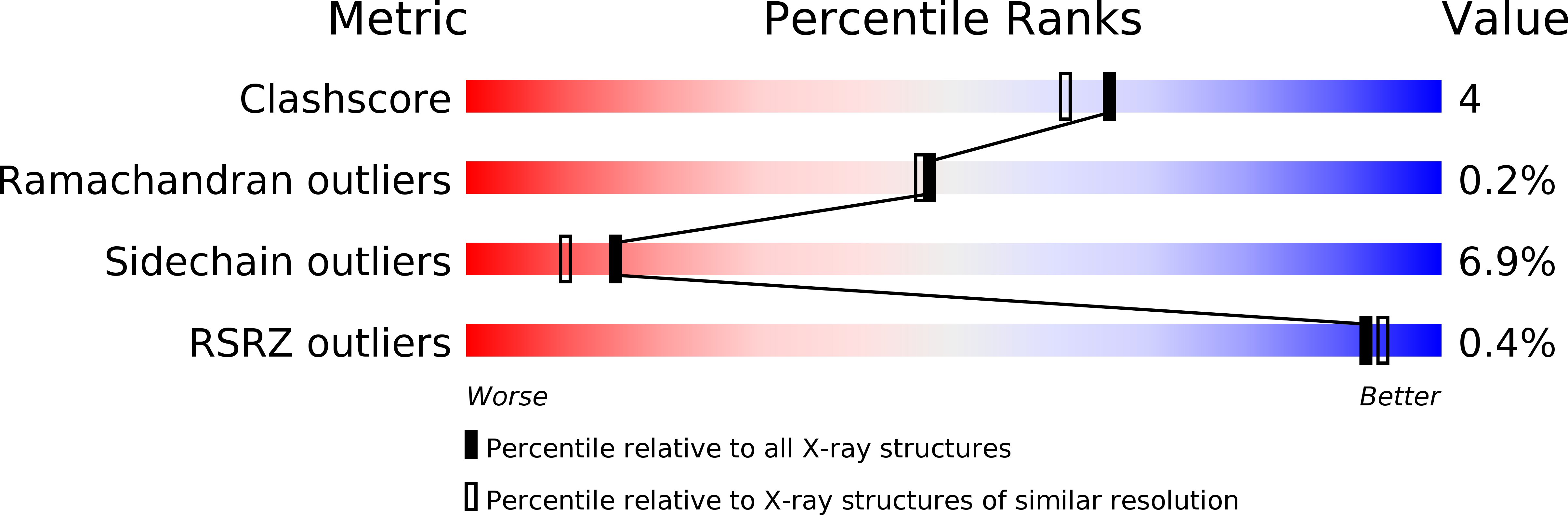
Resolution:
2.16 Å
R-Value Work:
0.17
R-Value Observed:
0.17
Space Group:
I 2 2 2
Macromolecular Entities
Structures with similar UniProt ID
Protein Blast
Polymer Type:polypeptide(L)
Molecule:NADH PEROXIDASE
Gene (Uniprot):npr
Chain IDs:A
Chain Length:447
Number of Molecules:1
Biological Source:Enterococcus faecalis
Modified Residue
Compound ID Chain ID Parent Comp ID Details 2D Image
OCS A CYS CYSTEINESULFONIC ACID
Ligand Molecules
Primary Citation
Structure of NADH peroxidase from Streptococcus faecalis 10C1 refined at 2.16 A resolution.
J.Mol.Biol. 221 1325 1344 (1991)
PMID: 1942054

Abstact

The crystal structure of NADH peroxidase (EC 1.11.1.1) from Streptococcus faecalis 10C1 (Enterococcus faecalis) has been refined to a resolution of 2.16 A using the simulated annealing method. The final crystallographic R-factor is 17.7% for all data in the resolution range 7 to 2.16 A. The standard deviations are 0.015 A in bond lengths and 3.0 degrees in bond angles for the final model, which includes all 447 amino acid residues, one FAD and 369 water molecules. The enzyme is a symmetrical tetramer with point group D2; the symmetry is crystallographic. The redox center of the enzyme consists of FAD and a cysteine (Cys42), which forms a sulfenic acid (Cys-SOH) in its oxidized state. A histidine (His10) close to Cys42 is likely to act as an active-site base. In the analyzed crystal, the enzyme was in a non-native oxidation state with Cys42 oxidized to a sulfonic acid Cys-SO3H. The chain fold of NADH peroxidase is similar to those of disulfide oxidoreductases. A comparison with glutathione reductase, a representative of this enzyme family, is given.

Legend

Protein

Chemical

Disease

Primary Citation of related structures