Deposition Date
1998-12-17
Release Date
1998-12-23
Last Version Date
2024-11-20
Entry Detail
PDB ID:
1NPL
Keywords:
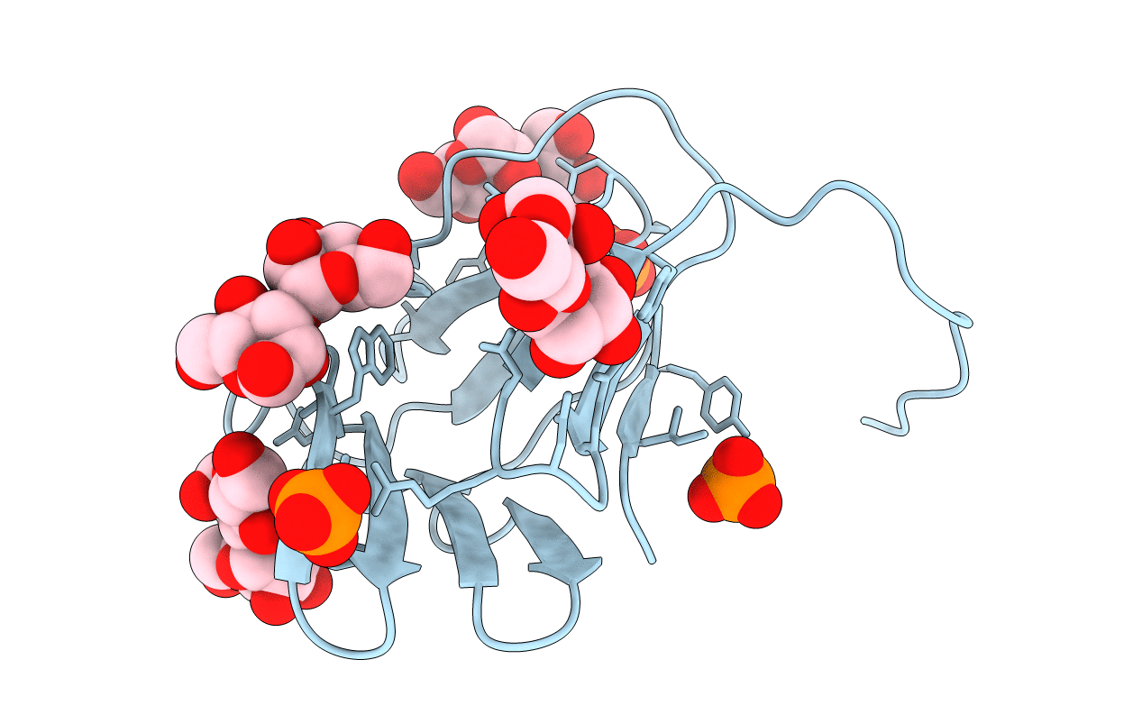
Title:
MANNOSE-SPECIFIC AGGLUTININ (LECTIN) FROM DAFFODIL (NARCISSUS PSEUDONARCISSUS) BULBS IN COMPLEX WITH MANNOSE-ALPHA1,3-MANNOSE
Biological Source:
Source Organism(s):
Narcissus pseudonarcissus (Taxon ID: 39639)
Method Details:
Experimental Method:
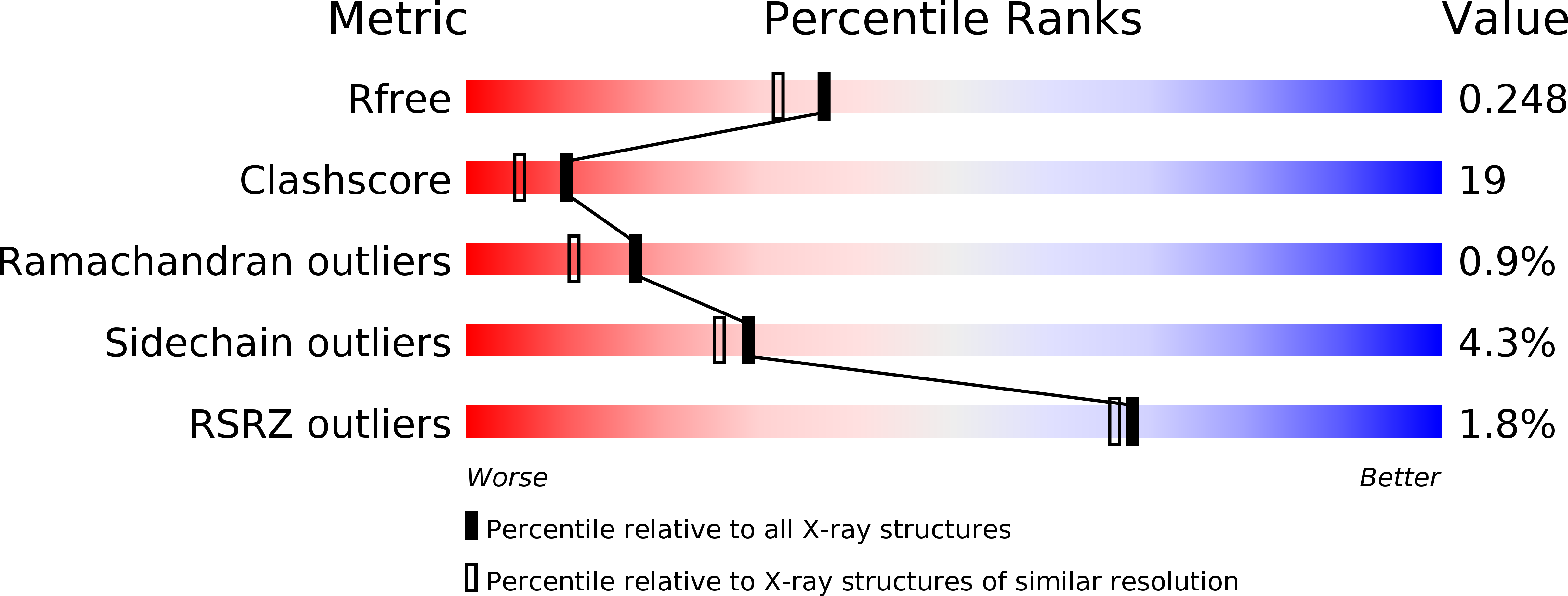
Resolution:
2.00 Å
R-Value Free:
0.26
R-Value Work:
0.18
Space Group:
C 2 2 2