Deposition Date
2003-01-17
Release Date
2003-05-13
Last Version Date
2023-08-16
Entry Detail
PDB ID:
1NPH
Keywords:
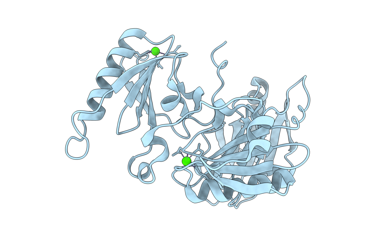
Title:
Gelsolin Domains 4-6 in Active, Actin Free Conformation Identifies Sites of Regulatory Calcium Ions
Biological Source:
Source Organism(s):
Mus musculus (Taxon ID: 10090)
Expression System(s):
Method Details:
Experimental Method:
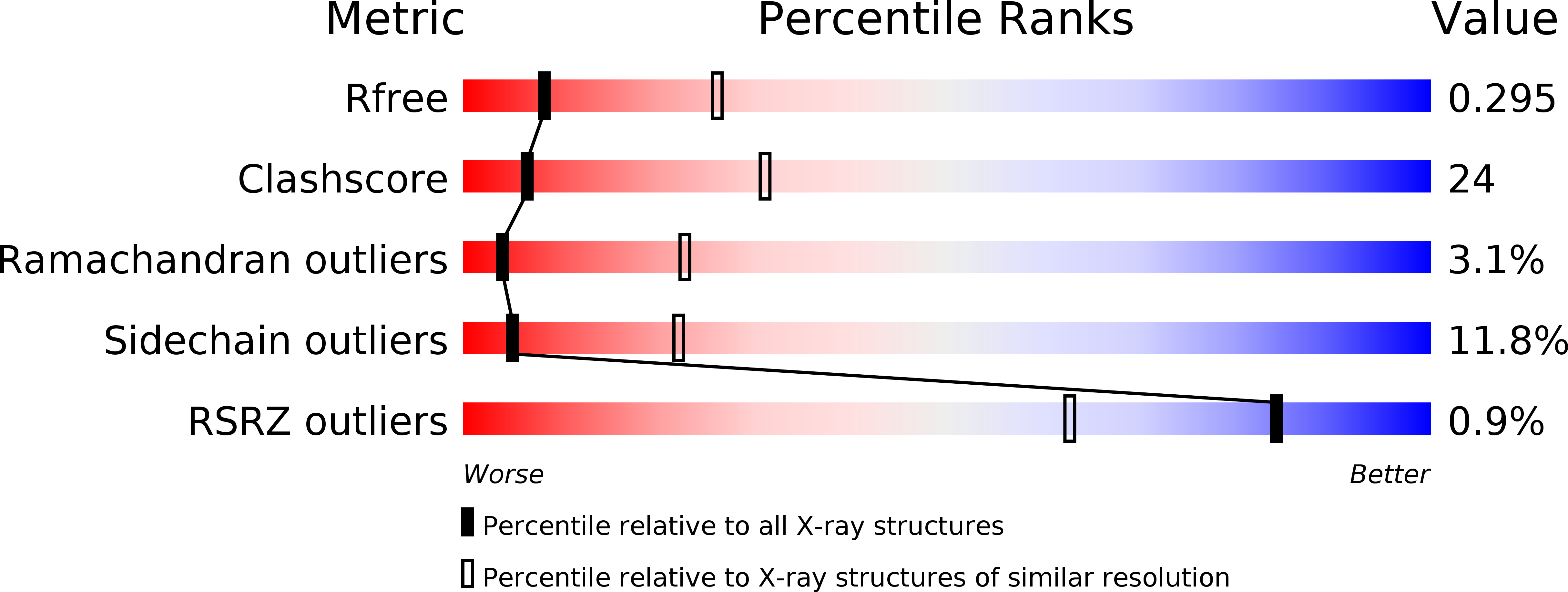
Resolution:
3.00 Å
R-Value Free:
0.29
R-Value Work:
0.24
Space Group:
P 41 21 2