1NOH image
Deposition Date 2003-01-16
Release Date 2003-07-01
Last Version Date 2023-08-16
Entry Detail
PDB ID:
1NOH
Keywords:
Title:
The structure of bacteriophage phi29 scaffolding protein gp7 after prohead assembly
Biological Source:
Source Organism:
Host Organism:
Method Details:
Experimental Method:
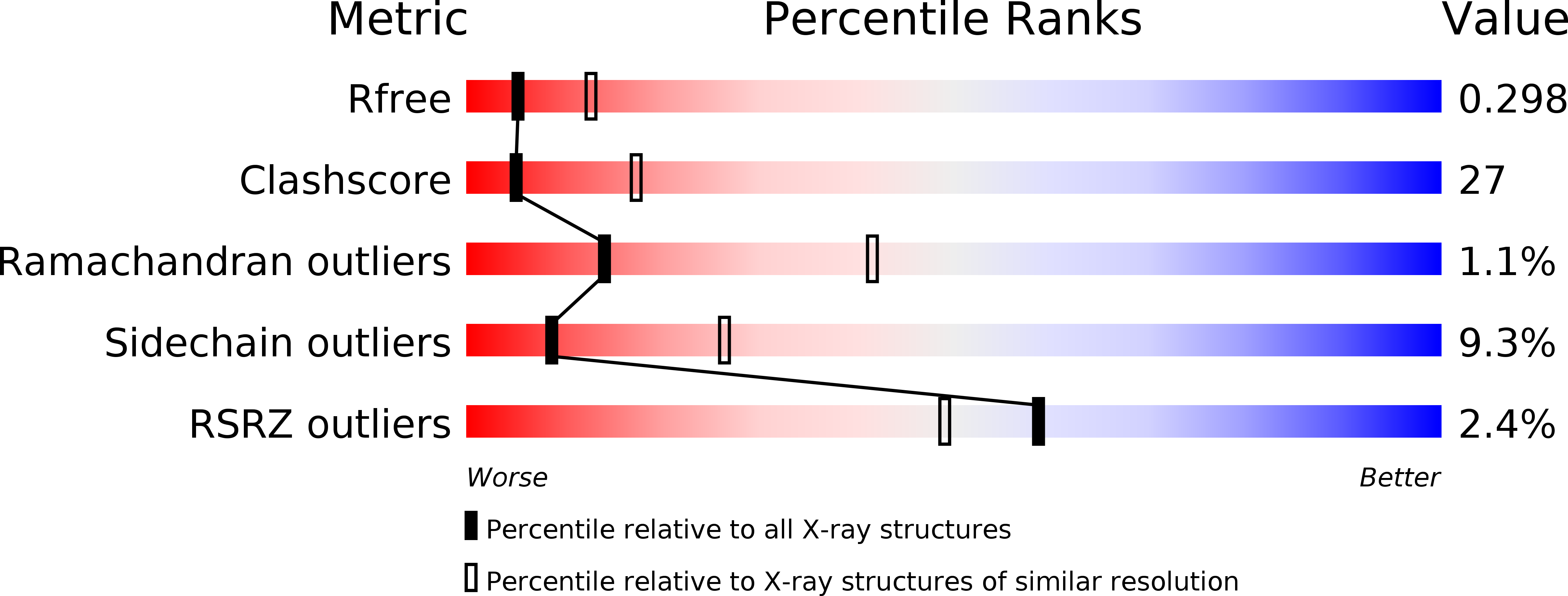
Resolution:
2.80 Å
R-Value Free:
0.29
R-Value Work:
0.25
R-Value Observed:
0.25
Space Group:
C 2 2 21
Macromolecular Entities
Structures with similar UniProt ID
Protein Blast
Polymer Type:polypeptide(L)
Molecule:HEAD MORPHOGENESIS PROTEIN
Gene (Uniprot):7
Chain IDs:A, B, C, D
Chain Length:97
Number of Molecules:4
Biological Source:Bacillus phage phi29
Ligand Molecules
Primary Citation
Bacteriophage f29 scaffolding protein gp7 before and after prohead assembly
Nat.Struct.Biol. 10 572 576 (2003)
PMID: 12778115 DOI: 10.1038/nsb939

Abstact

Three-dimensional structures of the double-stranded DNA bacteriophage phi29 scaffolding protein (gp7) before and after prohead assembly have been determined at resolutions of 2.2 and 2.8 A, respectively. Both structures are dimers that resemble arrows, with a four-helix bundle composing the arrowhead and a coiled coil forming the tail. The structural resemblance of gp7 to the yeast transcription factor GCN4 suggests a DNA-binding function that was confirmed by native gel electrophoresis. DNA binding to gp7 may have a role in mediating the structural transition from prohead to mature virus and scaffold release. A cryo-EM analysis indicates that gp7 is arranged inside the capsid as a series of concentric shells. The position of the higher density features in these shells correlates with the positions of hexamers in the equatorial region of the capsid, suggesting that gp7 may regulate formation of the prolate head through interactions with these hexamers.

Legend

Protein

Chemical

Disease

Primary Citation of related structures
Feedback Form
Name
Email
Institute
Feedback