1NNS image
Deposition Date 2003-01-14
Release Date 2003-03-11
Last Version Date 2024-10-30
Entry Detail
PDB ID:
1NNS
Keywords:
Title:
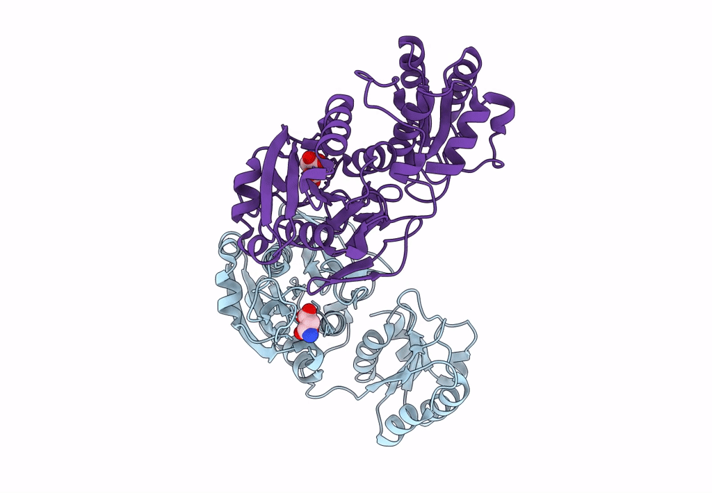
L-asparaginase of E. coli in C2 space group and 1.95 A resolution
Biological Source:
Source Organism:
Escherichia coli (Taxon ID: 562)
Method Details:
Experimental Method:
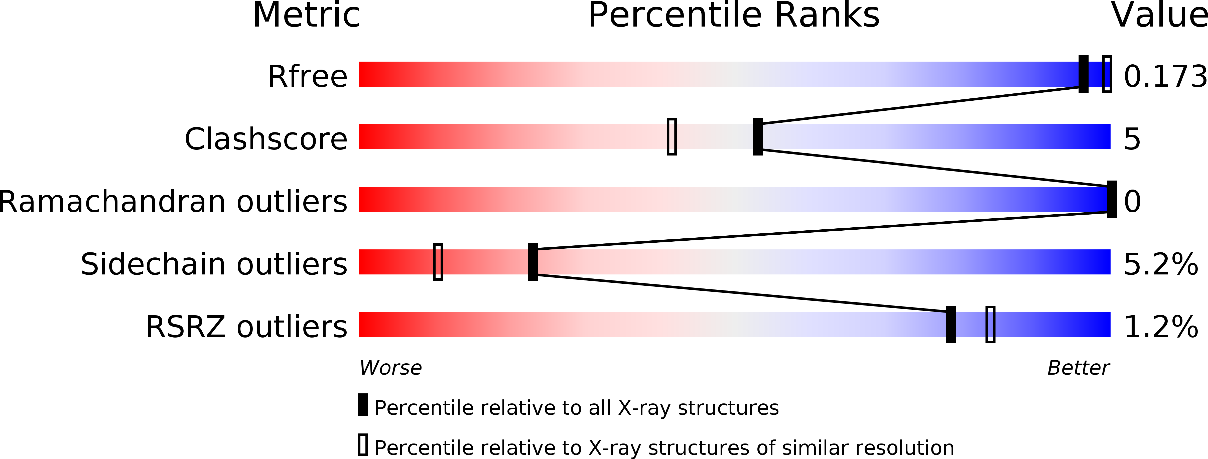
Resolution:
1.95 Å
R-Value Free:
0.17
R-Value Work:
0.12
R-Value Observed:
0.13
Space Group:
C 1 2 1
Macromolecular Entities
Polymer Type:polypeptide(L)
Molecule:L-asparaginase II
Gene (Uniprot):ansB
Chain IDs:A, B
Chain Length:326
Number of Molecules:2
Biological Source:Escherichia coli
Ligand Molecules
Primary Citation
Structural comparison of Escherichia coli L-asparaginase in two monoclinic space groups.
Acta Crystallogr.,Sect.D 59 416 422 (2003)
PMID: 12595697 DOI: 10.1107/S0907444902021200

Abstact

The functional L-asparaginase from Escherichia coli is a homotetramer with a molecular weight of about 142 kDa. The X-ray structure of the enzyme, crystallized in a new form (space group C2) and refined to 1.95 A resolution, is compared with that of the previously determined crystal form (space group P2(1)). The asymmetric unit of the new crystal form contains an L-asparaginase dimer instead of the tetramer found in the previous crystal form. It is found that crystal contacts practically do not affect the conformation of the protein. It is shown that subunit C of the tetrameric form is in a conformation which is systematically different from that of all other subunits in both crystal forms. Major conformational differences are confined to the lid loop (residues 14-27). In addition, the stability of this globular protein is analyzed in terms of the interactions between hydrophobic parts of the subunits.

Legend

Protein

Chemical

Disease

Primary Citation of related structures