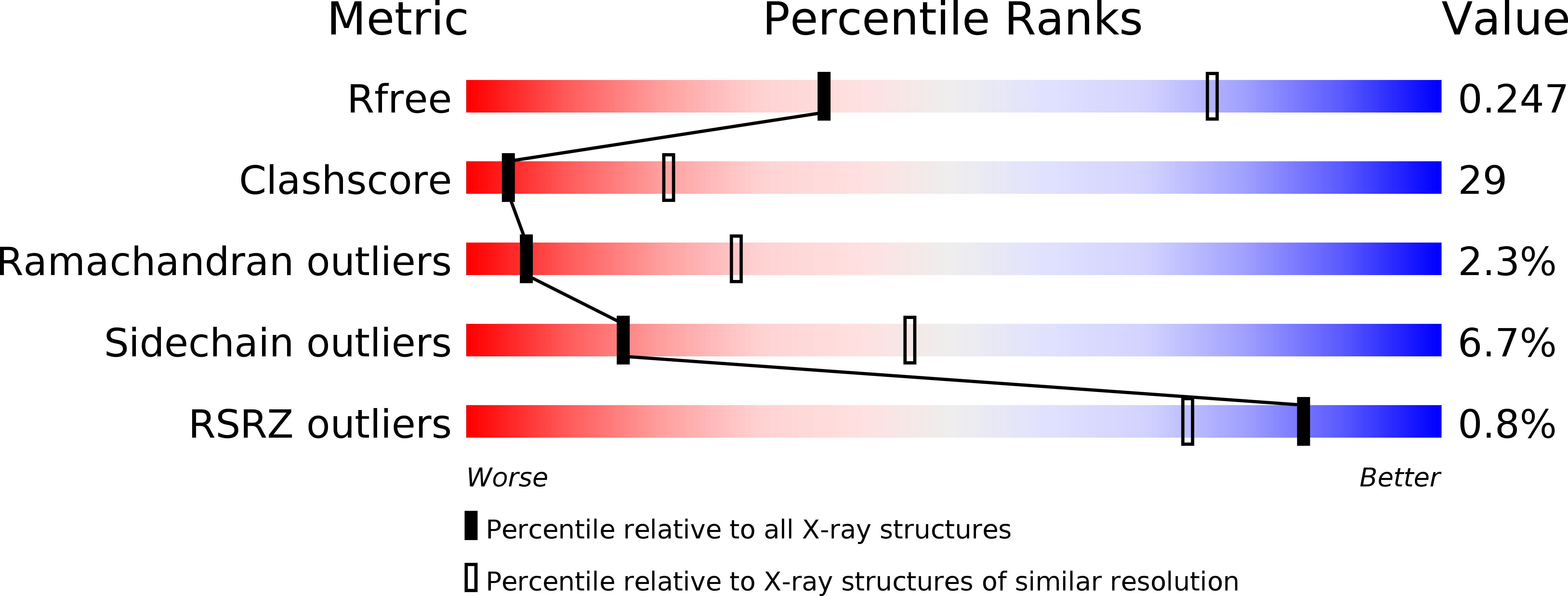
1NNE image
Deposition Date 2003-01-13
Release Date 2003-05-20
Last Version Date 2024-02-14
Entry Detail
PDB ID:
1NNE
Title:
Crystal Structure of the MutS-ADPBeF3-DNA complex
Biological Source:
Source Organism:
Method Details:
Experimental Method:
Resolution:
3.11 Å
R-Value Free:
0.25
R-Value Work:
0.20
R-Value Observed:
0.20
Space Group:
P 21 21 21
Macromolecular Entities
Structures with similar UniProt ID
Protein Blast
Polymer Type:polypeptide(L)
Molecule:DNA Mismatch Repair protein MutS
Gene (Uniprot):mutS
Chain IDs:C (auth: A), D (auth: B)
Chain Length:765
Number of Molecules:2
Biological Source:Thermus aquaticus
Primary Citation
Crystal structure and biochemical analysis of the MutS-ADP-Beryllium Fluoride complex suggests a conserved mechanism for ATP interactions in mismatch repair
J.Biol.Chem. 278 16088 16094 (2003)
PMID: 12582174 DOI: 10.1074/jbc.M213193200

Abstact

During mismatch repair ATP binding and hydrolysis activities by the MutS family proteins are important for both mismatch recognition and for transducing mismatch recognition signals to downstream repair factors. Despite intensive efforts, a MutS.ATP.DNA complex has eluded crystallographic analysis. Searching for ATP analogs that strongly bound to Thermus aquaticus (Taq) MutS, we found that ADP.beryllium fluoride (ABF), acted as a strong inhibitor of several MutS family ATPases. Furthermore, ABF promoted the formation of a ternary complex containing the Saccharomyces cerevisiae MSH2.MSH6 and MLH1.PMS1 proteins bound to mismatch DNA but did not promote dissociation of MSH2.MSH6 from mismatch DNA. Crystallographic analysis of the Taq MutS.DNA.ABF complex indicated that although this complex was very similar to that of MutS.DNA.ADP, both ADP.Mg(2+) moieties in the MutS. DNA.ADP structure were replaced by ABF. Furthermore, a disordered region near the ATP-binding pocket in the MutS B subunit became traceable, whereas the equivalent region in the A subunit that interacts with the mismatched nucleotide remained disordered. Finally, the DNA binding domains of MutS together with the mismatched DNA were shifted upon binding of ABF. We hypothesize that the presence of ABF is communicated between the two MutS subunits through the contact between the ordered loop and Domain III in addition to the intra-subunit helical lever arm that links the ATPase and DNA binding domains.

Legend

Protein

Chemical

Disease

Primary Citation of related structures