1NL0 image
Entry Detail
PDB ID:
1NL0
Keywords:
Title:
Crystal structure of human factor IX Gla domain in complex of an inhibitory antibody, 10C12
Biological Source:
Source Organism:
Host Organism:
PDB Version:
Deposition Date:
2003-01-06
Release Date:
2004-01-20
Method Details:
Experimental Method:
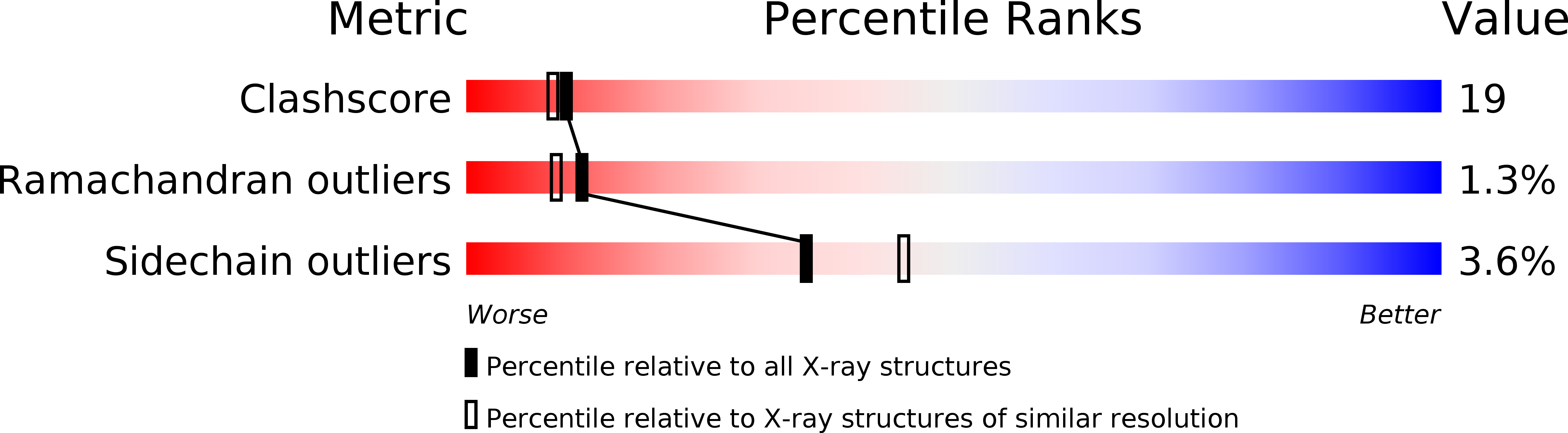
Resolution:
2.20 Å
R-Value Free:
0.27
R-Value Work:
0.23
R-Value Observed:
0.23
Space Group:
C 1 2 1
Macromolecular Entities
Polymer Type:polypeptide(L)
Description:factor IX
Chain IDs:C (auth: G)
Chain Length:51
Number of Molecules:1
Biological Source:
Polymer Type:polypeptide(L)
Description:anti-factor IX antibody, 10C12, chain H
Chain IDs:B (auth: H)
Chain Length:224
Number of Molecules:1
Biological Source:Homo sapiens
Polymer Type:polypeptide(L)
Description:anti-factor IX antibody, 10C12, chain L
Chain IDs:A (auth: L)
Chain Length:213
Number of Molecules:1
Biological Source:Homo sapiens
Modified Residue
Compound ID Chain ID Parent Comp ID Details 2D Image
CGU C GLU GAMMA-CARBOXY-GLUTAMIC ACID
Primary Citation
Crystal Structure of the Calcium-stabilized Human Factor IX Gla Domain Bound to a Conformation-specific Anti-factor IX Antibody.
J.Biol.Chem. 279 14338 14346 (2004)
PMID: 14722079 DOI: 10.1074/jbc.M314011200

Abstact

The binding of Factor IX to membranes during blood coagulation is mediated by the N-terminal gamma-carboxyglutamic acid-rich (Gla) domain, a membrane-anchoring domain found on vitamin K-dependent blood coagulation and regulatory proteins. Conformation-specific anti-Factor IX antibodies are directed at the calcium-stabilized Gla domain and interfere with Factor IX-membrane interaction. One such antibody, 10C12, recognizes the calcium-stabilized form of the Gla domain of Factor IX. We prepared the fully carboxylated Gla domain of Factor IX by solid phase peptide synthesis and crystallized Factor IX-(1-47) in complex with Fab fragments of the 10C12 antibody. The overall structure of the Gla domain in the Factor IX-(1-47)-antibody complex at 2.2 A is similar to the structure of the Factor IX Gla domain in the presence of calcium ions as determined by NMR spectroscopy (Freedman, S. J., Furie, B. C., Furie, B., and Baleja, J. D. (1995) Biochemistry 34, 12126-12137) and by x-ray crystallography (Shikamoto, Y., Morita, T., Fujimoto, Z., and Mizuno, H. (2003) J. Biol. Chem. 278, 24090-24094). The complex structure shows that the complementarity determining region loops of the 10C12 antibody form a hydrophobic pocket to accommodate the hydrophobic patch of the Gla domain consisting of Leu-6, Phe-9, and Val-10. Polar interactions also play an important role in the antibody-antigen recognition. Furthermore, the calcium coordination network of the Factor IX Gla domain is different than in Gla domain structures of other vitamin K-dependent proteins. We conclude that this antibody is directed at the membrane binding site in the omega loop of Factor IX and blocks Factor IX function by inhibiting its interaction with membranes.

Legend

Protein

Chemical

Disease

Primary Citation of related structures