Deposition Date
2003-01-05
Release Date
2003-02-11
Last Version Date
2023-08-16
Entry Detail
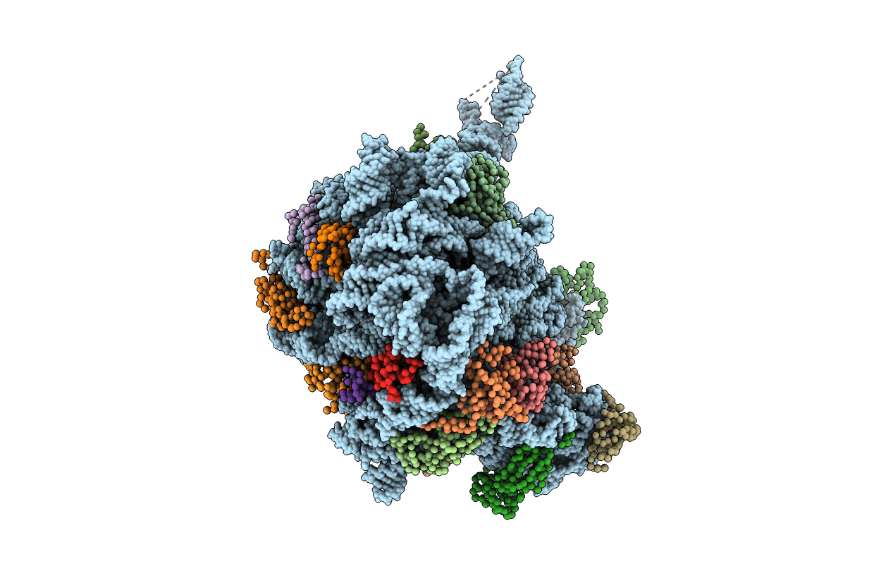
PDB ID:
1NKW
Keywords:
Title:
Crystal Structure Of The Large Ribosomal Subunit From Deinococcus Radiodurans
Biological Source:
Source Organism(s):
Deinococcus radiodurans (Taxon ID: 1299)
Method Details:
Experimental Method:
Resolution:
3.10 Å
R-Value Free:
0.27
R-Value Work:
0.24
Space Group:
I 2 2 2