Deposition Date
2003-01-02
Release Date
2003-06-10
Last Version Date
2023-08-16
Entry Detail
PDB ID:
1NJS
Keywords:
Title:
human GAR Tfase in complex with hydrolyzed form of 10-trifluoroacetyl-5,10-dideaza-acyclic-5,6,7,8-tetrahydrofolic acid
Biological Source:
Source Organism(s):
Homo sapiens (Taxon ID: 9606)
Expression System(s):
Method Details:
Experimental Method:
Resolution:
1.98 Å
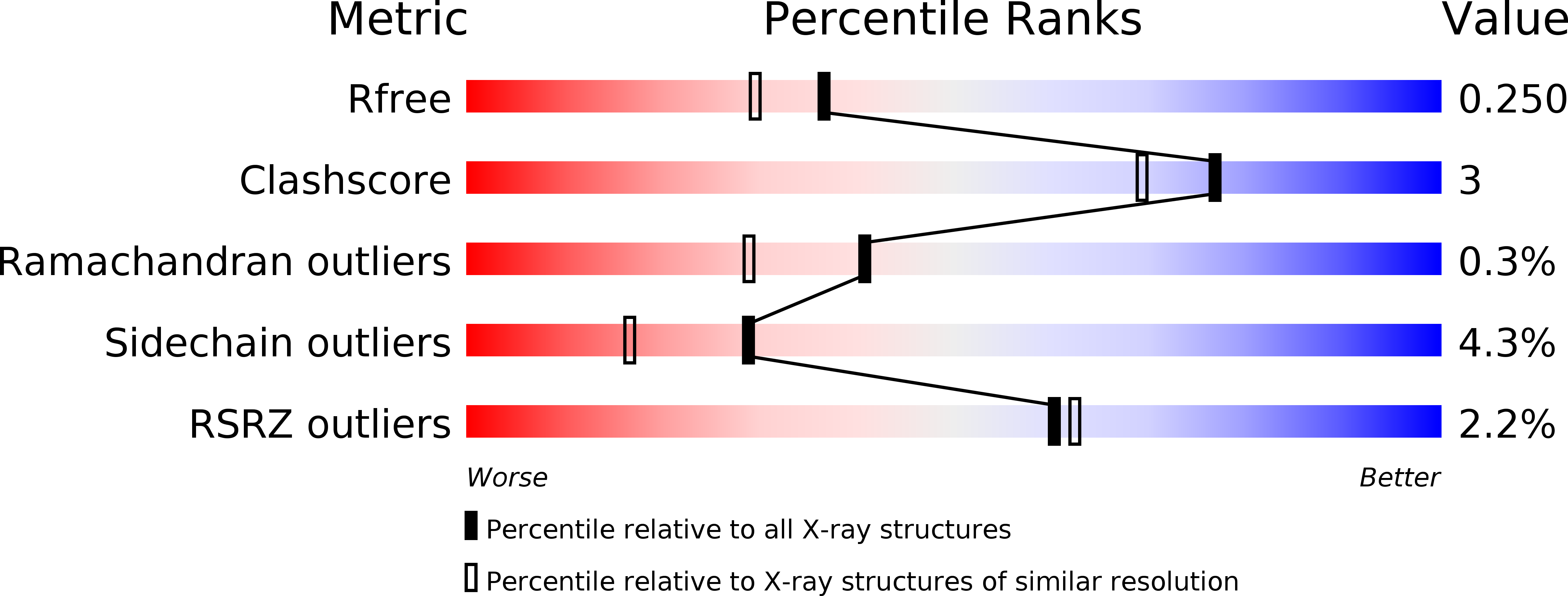
R-Value Free:
0.24
R-Value Work:
0.22
R-Value Observed:
0.22
Space Group:
P 31 2 1