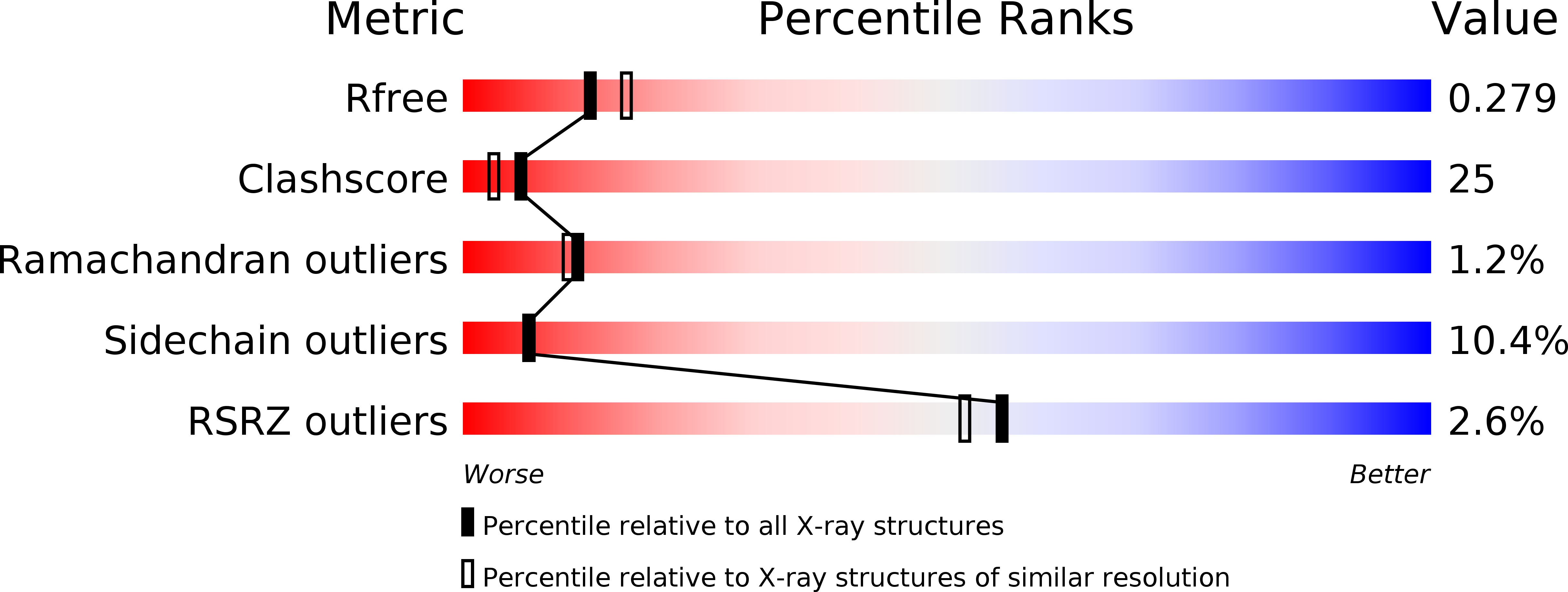
1NJJ image
Deposition Date 2002-12-31
Release Date 2003-08-26
Last Version Date 2023-08-16
Entry Detail
PDB ID:
1NJJ
Keywords:
Title:
Crystal structure determination of T. brucei ornithine decarboxylase bound to D-ornithine and to G418
Biological Source:
Source Organism:
Host Organism:
Method Details:
Experimental Method:
Resolution:
2.45 Å
R-Value Free:
0.28
R-Value Work:
0.26
Space Group:
P 1 21 1
Macromolecular Entities
Structures with similar UniProt ID
Protein Blast
Polymer Type:polypeptide(L)
Molecule:ornithine decarboxylase
Chain IDs:A, B, C, D
Chain Length:425
Number of Molecules:4
Biological Source:Trypanosoma brucei gambiense
Primary Citation
X-ray Structure Determination of Trypanosoma brucei Ornithine Decarboxylase Bound to D-Ornithine and to G418: INSIGHTS INTO SUBSTRATE BINDING AND ODC CONFORMATIONAL FLEXIBILITY.
J.Biol.Chem. 278 22037 22043 (2003)
PMID: 12672797 DOI: 10.1074/jbc.M300188200

Abstact

Ornithine decarboxylase (ODC) is a pyridoxal 5'-phosphate (PLP)-dependent enzyme that catalyzes the rate-determining step in the biosynthesis of polyamines. ODC is a proven drug target to treat African sleeping sickness. The x-ray crystal structure of Trypanosoma brucei ODC in complex with d-ornithine (d-Orn), a substrate analog, and G418 (Geneticin), a weak non-competitive inhibitor, was determined to 2.5-A resolution. d-Orn forms a Schiff base with PLP, and the side chain is in a similar position to that observed for putrescine and alpha-difluoromethylornithine in previous T. brucei ODC structures. The d-Orn carboxylate is positioned on the solvent-exposed side of the active site (si face of PLP), and Gly-199, Gly-362, and His-197 are the only residues within 4.2 A of this moiety. This structure confirms predictions that the carboxylate of d-Orn binds on the si face of PLP, and it supports a model in which the carboxyl group of the substrate l-Orn would be buried on the re face of the cofactor in a pocket that includes Phe-397, Tyr-389, Lys-69 (methylene carbons), and Asp-361. Electron density for G418 was observed at the boundary between the two domains within each ODC monomer. A ten-amino acid loop region (392-401) near the 2-fold axis of the dimer interface, which contributes several residues that form the active site, is disordered in this structure. The disordering of residues in the active site provides a potential mechanism for inhibition by G418 and suggests that allosteric inhibition from this site is feasible.

Legend

Protein

Chemical

Disease

Primary Citation of related structures