Deposition Date
2002-12-19
Release Date
2003-01-07
Last Version Date
2024-10-30
Entry Detail
PDB ID:
1NHE
Keywords:
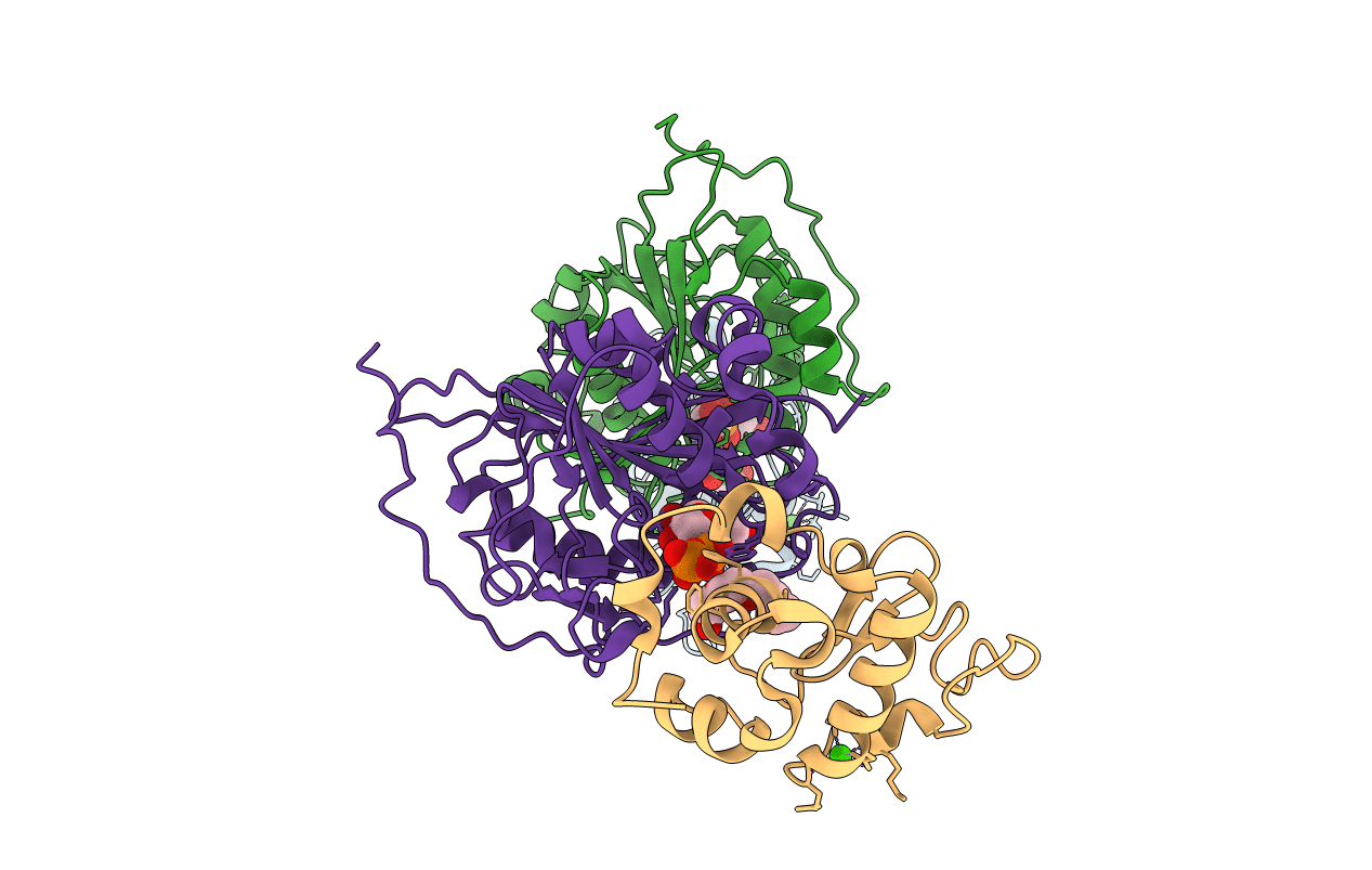
Title:
Crystal structure of Lactose synthase complex with UDP
Biological Source:
Source Organism(s):
Mus musculus (Taxon ID: 10090)
Bos taurus (Taxon ID: 9913)
Bos taurus (Taxon ID: 9913)
Expression System(s):
Method Details:
Experimental Method:
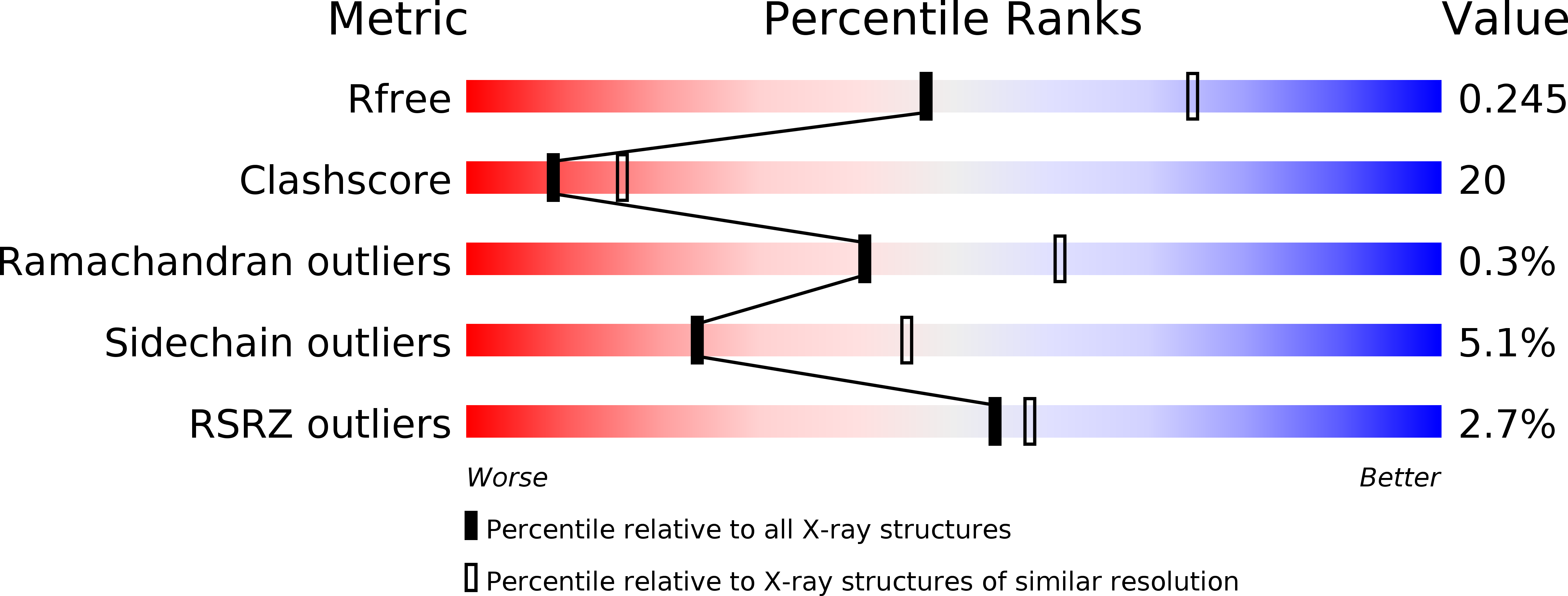
Resolution:
2.50 Å
R-Value Free:
0.25
R-Value Work:
0.19
R-Value Observed:
0.19
Space Group:
P 1 21 1