1NH0 image
Deposition Date 2002-12-18
Release Date 2004-04-13
Last Version Date 2023-08-16
Entry Detail
PDB ID:
1NH0
Title:
1.03 A structure of HIV-1 protease: inhibitor binding inside and outside the active site
Biological Source:
Source Organism:
Host Organism:
Method Details:
Experimental Method:
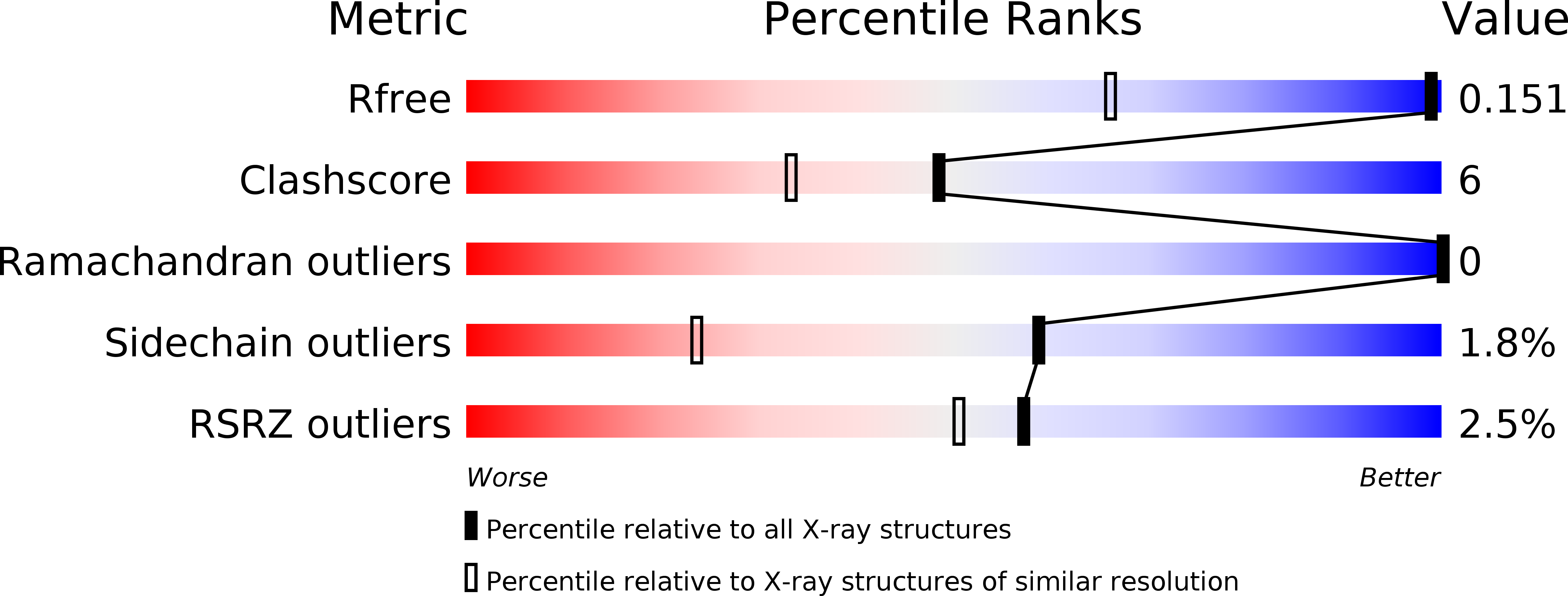
Resolution:
1.03 Å
R-Value Free:
0.16
R-Value Work:
0.13
R-Value Observed:
0.13
Space Group:
P 21 21 21
Macromolecular Entities
Polymer Type:polypeptide(L)
Molecule:PROTEASE RETROPEPSIN
Gene (Uniprot):gag-pol
Chain IDs:A, B
Chain Length:99
Number of Molecules:2
Biological Source:Human immunodeficiency virus 1
Peptide-like Molecules
PRD_000401
Primary Citation
A Phenylnorstatine Inhibitor Binding to HIV-1 Protease: Geometry, Protonation, and Subsite-Pocket Interactions Analyzed at Atomic Resolution
J.Med.Chem. 47 2030 2036 (2004)
PMID: 15056001 DOI: 10.1021/jm031105q

Abstact

The X-ray structure of a complex of HIV-1 protease (PR) with a phenylnorstatine inhibitor Z-Pns-Phe-Glu-Glu-NH(2) has been determined at 1.03 A, the highest resolution so far reported for any HIV PR complex. The inhibitor shows subnanomolar K(i) values for both the wild-type PR and the variant representing one of the most common mutations linked to resistance development. The structure comprising the phenylnorstatine moiety of (2R,3S)-chirality displays a unique pattern of hydrogen bonding to the two catalytic aspartate residues. This high resolution makes it possible to assess the donor and acceptor relations of this hydrogen bonding and to indicate a proton shared by the two catalytic residues. A structural mechanism for the unimpaired inhibition of the protease Val82Ala mutant is also suggested, based on energy calculations and analyses.

Legend

Protein

Chemical

Disease

Primary Citation of related structures