1NG5 image
Deposition Date 2002-12-16
Release Date 2003-09-23
Last Version Date 2024-05-22
Entry Detail
PDB ID:
1NG5
Title:
2.0 A crystal structure of Staphylococcus aureus Sortase B
Biological Source:
Source Organism:
Method Details:
Experimental Method:
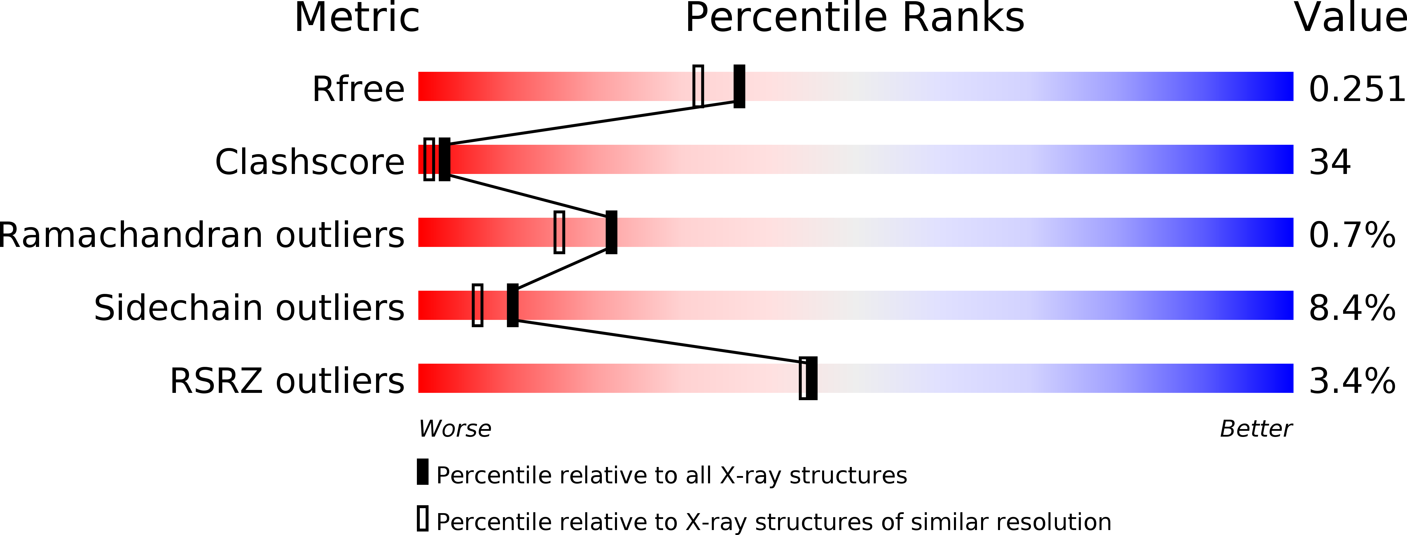
Resolution:
2.00 Å
R-Value Free:
0.24
R-Value Work:
0.23
R-Value Observed:
0.23
Space Group:
P 21 21 2
Macromolecular Entities
Polymer Type:polypeptide(L)
Molecule:sortase B
Chain IDs:A, B
Chain Length:215
Number of Molecules:2
Biological Source:Staphylococcus aureus subsp. aureus
Primary Citation
Structures of sortase B from Staphylococcus aureus and Bacillus anthracis reveal catalytic amino acid triad in the active site.
Structure 12 1147 1156 (2004)
PMID: 15242591 DOI: 10.1016/j.str.2004.06.001

Abstact

Surface proteins attached by sortases to the cell wall envelope of bacterial pathogens play important roles during infection. Sorting and attachment of these proteins is directed by C-terminal signals. Sortase B of S. aureus recognizes a motif NPQTN, cleaves the polypeptide after the Thr residue, and attaches the protein to pentaglycine cross-bridges. Sortase B of B. anthracis is thought to recognize the NPKTG motif, and attaches surface proteins to m-diaminopimelic acid cross-bridges. We have determined crystal structure of sortase B from B. anthracis and S. aureus at 1.6 and 2.0 A resolutions, respectively. These structures show a beta-barrel fold with alpha-helical elements on its outside, a structure thus far exclusive to the sortase family. A putative active site located on the edge of the beta-barrel is comprised of a Cys-His-Asp catalytic triad and presumably faces the bacterial cell surface. A putative binding site for the sorting signal is located nearby.

Legend

Protein

Chemical

Disease

Primary Citation of related structures