1NF4 image
Deposition Date 2002-12-13
Release Date 2003-04-01
Last Version Date 2024-04-03
Entry Detail
PDB ID:
1NF4
Title:
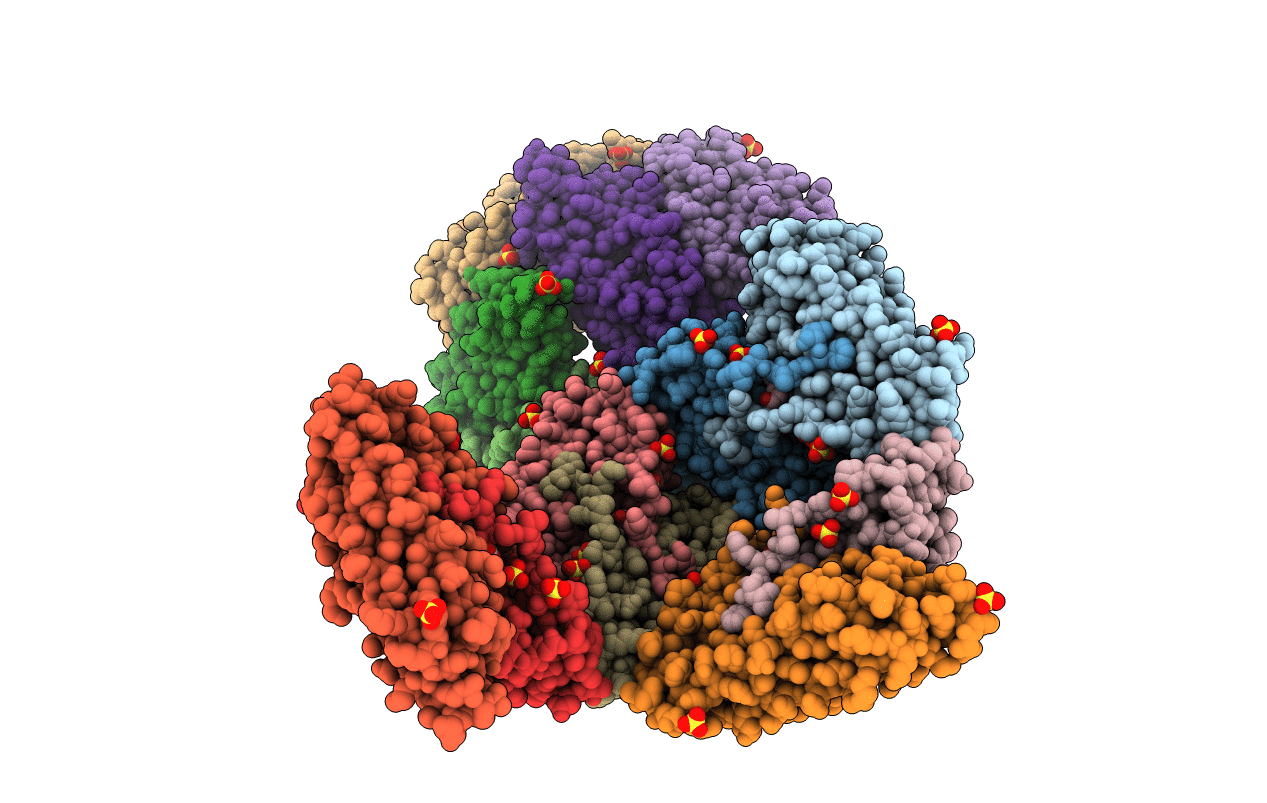
X-Ray Structure of the Desulfovibrio desulfuricans bacterioferritin: the diiron site in different states (reduced structure)
Biological Source:
Source Organism:
Method Details:
Experimental Method:
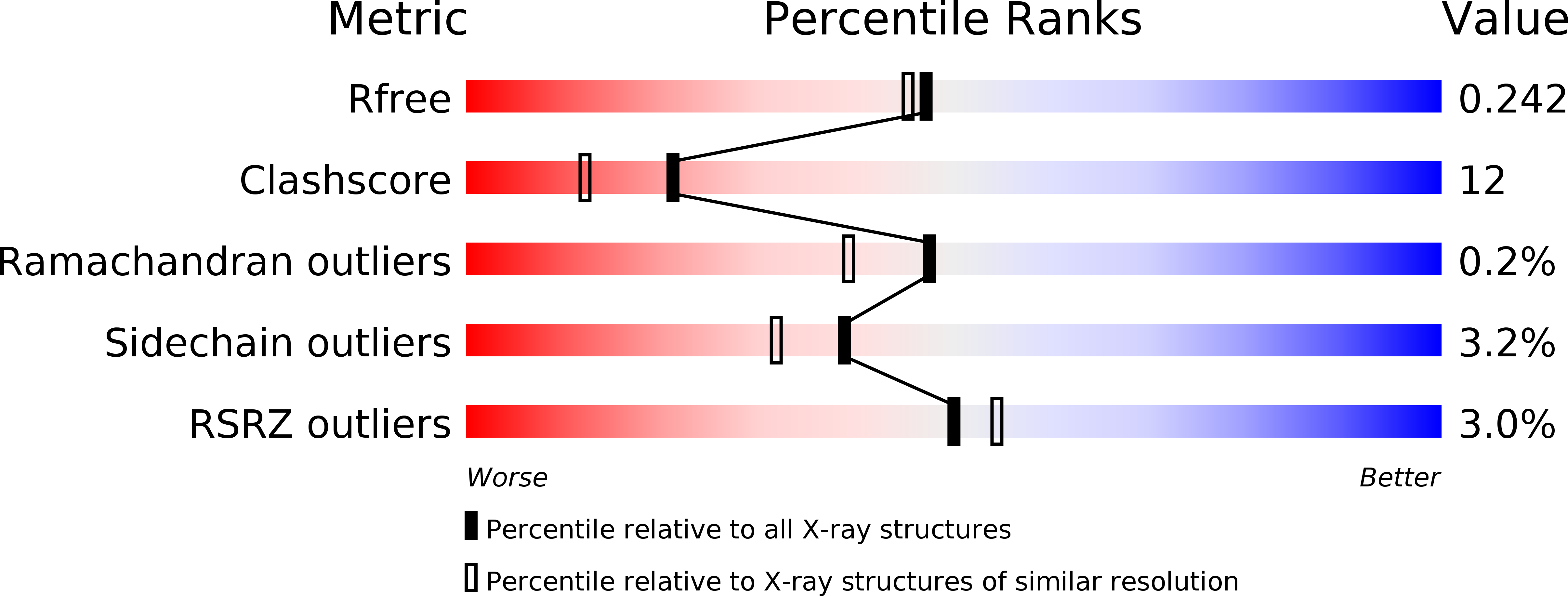
Resolution:
2.05 Å
R-Value Free:
0.27
R-Value Work:
0.23
R-Value Observed:
0.23
Space Group:
P 21 3
Macromolecular Entities
Polymer Type:polypeptide(L)
Molecule:bacterioferritin
Chain IDs:A, B, C, D, E, F, G, H, I, J, K, L, M, N, O, P
Chain Length:179
Number of Molecules:16
Biological Source:Desulfovibrio desulfuricans
Primary Citation
The nature of the di-iron site in the bacterioferritin from Desulfovibrio desulfuricans
NAT.STRUCT.BIOL. 10 285 290 (2003)
PMID: 12627224 DOI: 10.1038/nsb909

Abstact

The first crystal structure of a native di-iron center in an iron-storage protein (bacterio)ferritin is reported. The protein, isolated from the anaerobic bacterium Desulfovibrio desulfuricans, has the unique property of having Fe-coproporphyrin III as its heme cofactor. The three-dimensional structure of this bacterioferritin was determined in three distinct catalytic/redox states by X-ray crystallography (at 1.95, 2.05 and 2.35 A resolution), corresponding to different intermediates of the di-iron ferroxidase site. Conformational changes associated with these intermediates support the idea of a route for iron entry into the protein shell through a pore that passes through the di-iron center. Molecular surface and electrostatic potential calculations also suggest the presence of another ion channel, distant from the channels at the three- and four-fold axes proposed as points of entry for the iron atoms.

Legend

Protein

Chemical

Disease

Primary Citation of related structures