Deposition Date
2002-12-12
Release Date
2003-03-04
Last Version Date
2024-10-30
Entry Detail
PDB ID:
1NF3
Keywords:
Title:
Structure of Cdc42 in a complex with the GTPase-binding domain of the cell polarity protein, Par6
Biological Source:
Source Organism(s):
Homo sapiens (Taxon ID: 9606)
Mus musculus (Taxon ID: 10090)
Mus musculus (Taxon ID: 10090)
Expression System(s):
Method Details:
Experimental Method:
Resolution:
2.10 Å
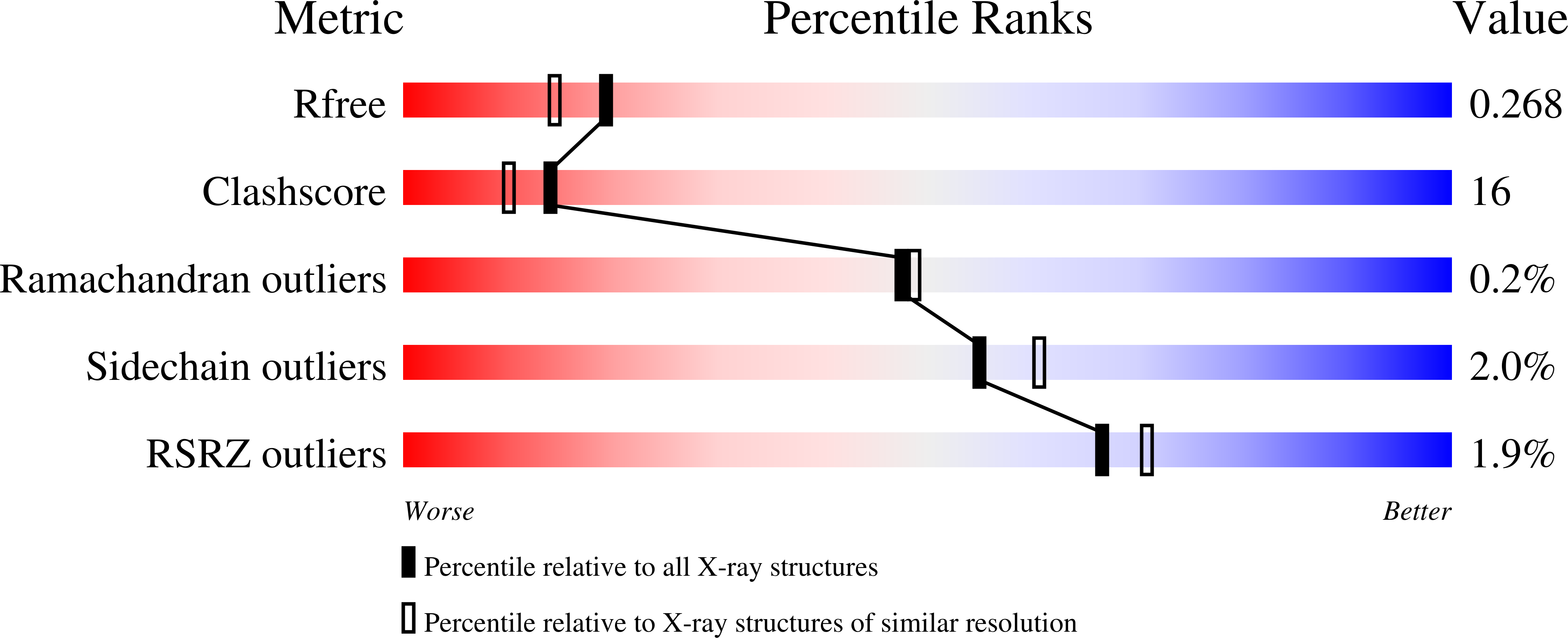
R-Value Free:
0.27
R-Value Work:
0.21
R-Value Observed:
0.21
Space Group:
P 1