1NEA image
Deposition Date 1992-09-22
Release Date 1993-10-31
Last Version Date 2024-10-30
Entry Detail
PDB ID:
1NEA
Keywords:
Title:
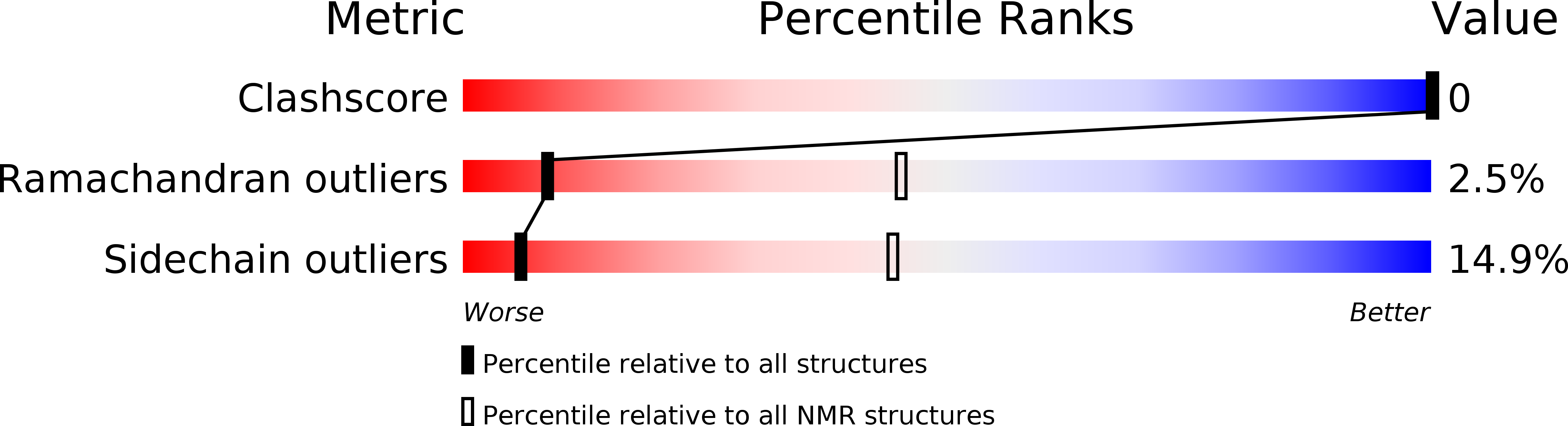
THREE-DIMENSIONAL SOLUTION STRUCTURE OF A CURAREMIMETIC TOXIN FROM NAJA NIGRICOLLIS VENOM: A PROTON NMR AND MOLECULAR MODELING STUDY
Biological Source:
Source Organism:
Method Details:
Experimental Method:
Conformers Submitted:
8
Macromolecular Entities
Polymer Type:polypeptide(L)
Molecule:TOXIN ALPHA
Chain IDs:A
Chain Length:61
Number of Molecules:1
Biological Source:Naja nigricollis
Ligand Molecules
Primary Citation
Three-dimensional solution structure of a curaremimetic toxin from Naja nigricollis venom: a proton NMR and molecular modeling study.
Biochemistry 31 11335 11347 (1992)
PMID: 1332755 DOI: 10.1021/bi00161a011

Abstact

The solution conformation of toxin alpha from Naja nigricollis (61 amino acids and four disulfides), a snake toxin which specifically blocks the activity of the nicotinic acetylcholine receptor (AcChoR), has been determined using nuclear magnetic resonance spectroscopy and molecular modeling. The solution structures were calculated using 409 distance and 73 dihedral angle restraints. The average atomic rms deviation between the eight refined structures and the mean structure is approximately 0.5 A for the backbone atoms. The overall folding of toxin alpha consists of three major loops which are stabilized by three disulfide bridges and one short C terminal loop stabilized by a fourth disulfide bridge. All the disulfides are grouped in the same region of the molecule, forming a highly constrained structure from which the loops protrude. As predicted, this structure appears to be very similar to the 1.4-A resolution crystal structure of another snake neurotoxin, namely, erabutoxin b from Laticauda semifasciata. The atomic rms deviation for the backbone atoms between the solution and crystal structures is approximately 1.7 A. The minor differences which are observed between the two structures are partly related to the deletion of one residue from the chain of toxin alpha. It is notable that, although the two toxins differ from each other by 16 amino acid substitutions, their side chains have an essentially similar spatial organization. However, most of the side chains which constitute the presumed AcChoR binding site for the curaremimetic toxins are poorly resolved in toxin alpha.(ABSTRACT TRUNCATED AT 250 WORDS)

Legend

Protein

Chemical

Disease

Primary Citation of related structures