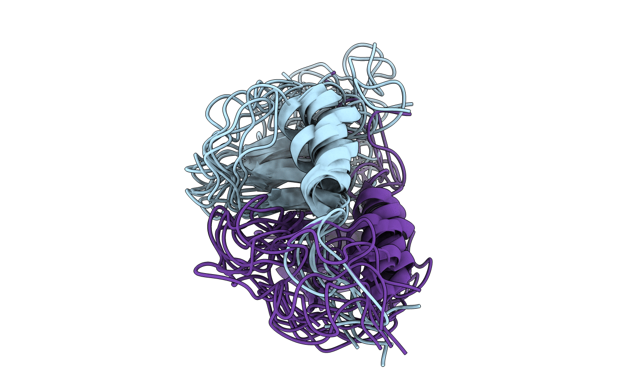
1NCV image
Deposition Date 1997-02-05
Release Date 1997-10-15
Last Version Date 2024-10-23
Entry Detail
PDB ID:
1NCV
Keywords:
Title:
DETERMINATION CC-CHEMOKINE MCP-3, NMR, 7 STRUCTURES
Biological Source:
Source Organism:
Homo sapiens (Taxon ID: 9606)
Host Organism:
Method Details:
Experimental Method:
Conformers Calculated:
50
Conformers Submitted:
7
Selection Criteria:
NOE VIOLATION
Macromolecular Entities
Structures with similar UniProt ID
Protein Blast
Polymer Type:polypeptide(L)
Molecule:MONOCYTE CHEMOATTRACTANT PROTEIN 3
Gene (Uniprot):CCL7
Chain IDs:A, B
Chain Length:76
Number of Molecules:2
Biological Source:Homo sapiens
Ligand Molecules
Primary Citation
Determination of the three-dimensional structure of CC chemokine monocyte chemoattractant protein 3 by 1H two-dimensional NMR spectroscopy.
Biochemistry 36 4412 4422 (1997)
PMID: 9109648 DOI: 10.1021/bi9627929

Abstact

MCP-3 is a beta chemokine consisting of 76 amino acid residues. It has been described to be involved in the activation of all leukocytic cells, activation mediated by the presence of multiple binding sites on the target cells. Its three-dimensional structure has been studied by making use of two-dimensional 1H NMR spectroscopy. MCP-3 exhibits the same monomeric structure as the other chemokines, i.e., a three-stranded antiparallel beta sheet covered on one face by an alpha helix. Although it belongs to the same subfamily as RANTES (Chung et al., 1995; Faitbrother et al., 1994) and hMIP-1beta (Lodi et al., 1994), the MCP-3 dimer is folded like IL-8 with the so-called alphabeta sandwich structural motif. Structural and sequence analysis gives clear indications suggesting that the other MCP chemokines may have the same quaternary structure, contrary to the other beta chemokines.

Legend

Protein

Chemical

Disease

Primary Citation of related structures