1NC1 image
Deposition Date 2002-12-04
Release Date 2003-11-25
Last Version Date 2023-08-16
Entry Detail
PDB ID:
1NC1
Keywords:
Title:
Crystal structure of E. coli MTA/AdoHcy nucleosidase complexed with 5'-methylthiotubercidin (MTH)
Biological Source:
Source Organism:
Escherichia coli (Taxon ID: 562)
Host Organism:
Method Details:
Experimental Method:
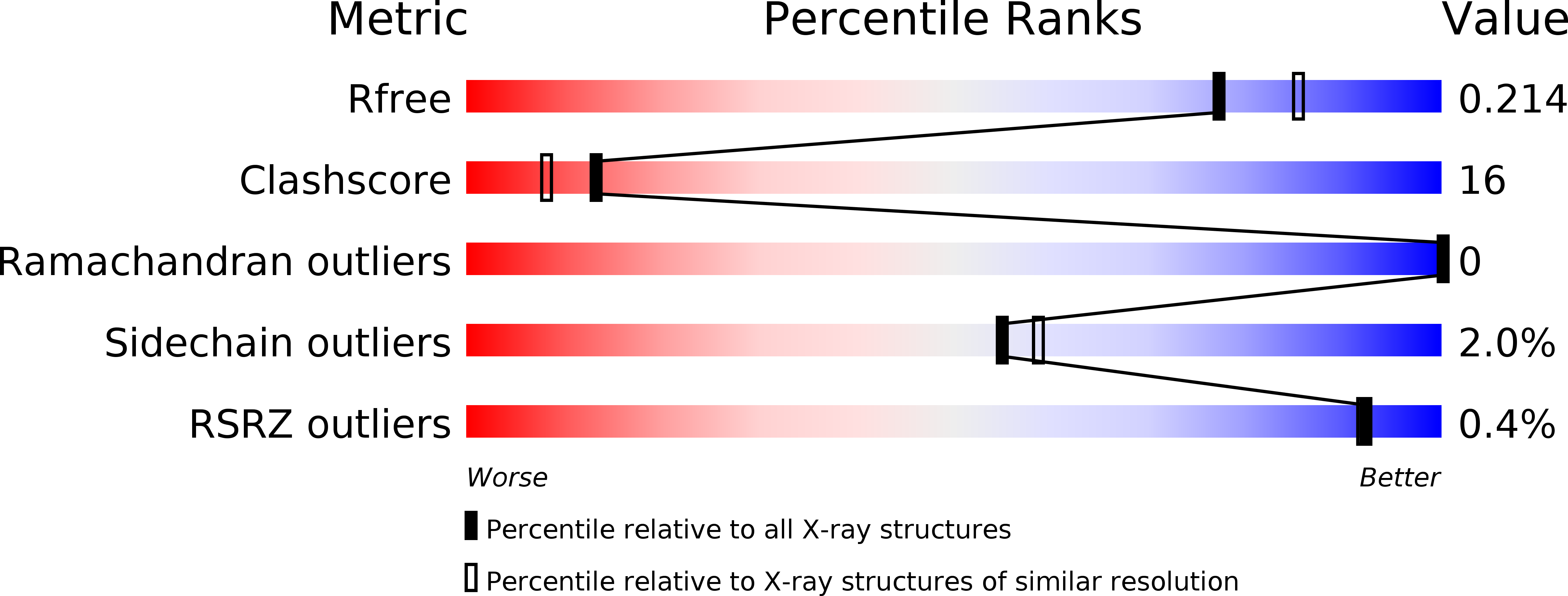
Resolution:
2.00 Å
R-Value Free:
0.22
R-Value Work:
0.19
Space Group:
P 21 21 21
Macromolecular Entities
Polymer Type:polypeptide(L)
Molecule:MTA/SAH nucleosidase
Chain IDs:A, B
Chain Length:242
Number of Molecules:2
Biological Source:Escherichia coli
Ligand Molecules
Primary Citation
Structure of Escherichia coli 5'-methylthioadenosine/ S-adenosylhomocysteine nucleosidase inhibitor complexes provide insight into the conformational changes required for substrate binding and catalysis.
J.Biol.Chem. 278 8761 8770 (2003)
PMID: 12496243 DOI: 10.1074/jbc.M210836200

Abstact

5'-Methylthioadenosine/S-adenosylhomocysteine (MTA/AdoHcy) nucleosidase is a key enzyme in a number of critical biological processes in many microbes. This nucleosidase catalyzes the irreversible hydrolysis of the N(9)-C(1') bond of MTA or AdoHcy to form adenine and the corresponding thioribose. The key role of the MTA/AdoHcy nucleosidase in biological methylation, polyamine biosynthesis, methionine recycling, and bacterial quorum sensing has made it an important antimicrobial drug target. The crystal structures of Escherichia coli MTA/AdoHcy nucleosidase complexed with the transition state analog, formycin A (FMA), and the nonhydrolyzable substrate analog, 5'-methylthiotubercidin (MTT) have been solved to 2.2- and 2.0-A resolution, respectively. These are the first MTA/AdoHcy nucleosidase structures to be solved in the presence of inhibitors. These structures clearly identify the residues involved in substrate binding and catalysis in the active site. Comparisons of the inhibitor complexes to the adenine-bound MTA/AdoHcy nucleosidase (Lee, J. E., Cornell, K. A., Riscoe, M. K., and Howell, P. L. (2001) Structure (Camb.) 9, 941-953) structure provide evidence for a ligand-induced conformational change in the active site and the substrate preference of the enzyme. The enzymatic mechanism has been re-examined.

Legend

Protein

Chemical

Disease

Primary Citation of related structures