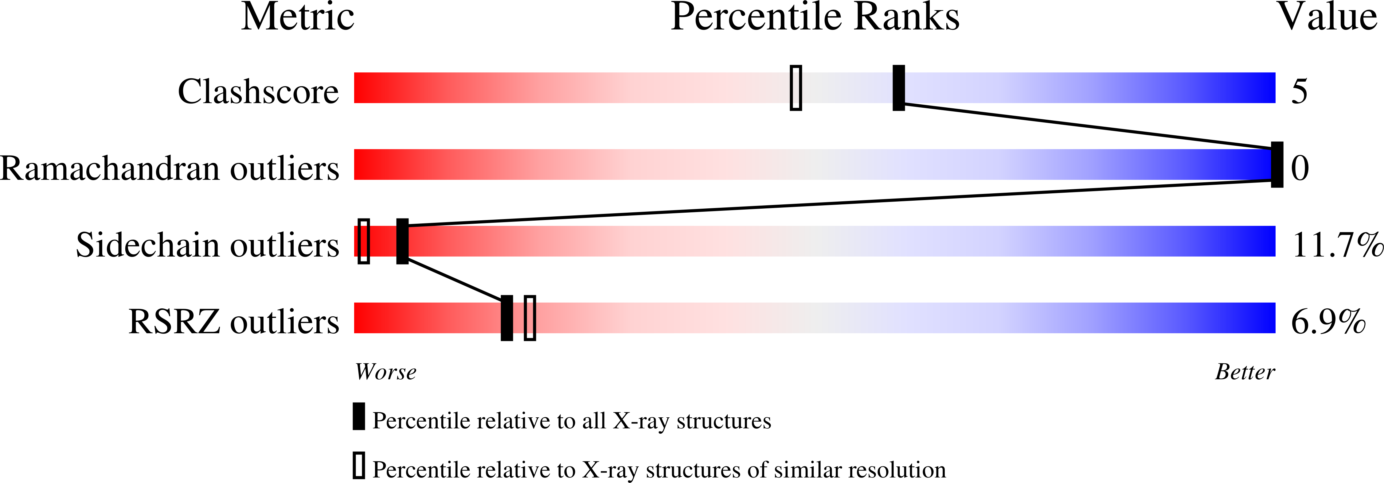
1NBX image
Deposition Date 2002-12-04
Release Date 2003-09-02
Last Version Date 2023-08-16
Entry Detail
PDB ID:
1NBX
Title:
Streptavidin Mutant Y43A at 1.70A Resolution
Biological Source:
Source Organism:
Host Organism:
Method Details:
Experimental Method:
Resolution:
1.70 Å
R-Value Free:
0.27
R-Value Work:
0.20
R-Value Observed:
0.21
Space Group:
P 1 21 1
Macromolecular Entities
Polymer Type:polypeptide(L)
Molecule:Streptavidin
Mutations:Y43A
Chain IDs:A, B, C, D
Chain Length:127
Number of Molecules:4
Biological Source:Streptomyces avidinii
Ligand Molecules
Primary Citation
Structural studies of hydrogen bonds in the high-affinity streptavidin-biotin complex: mutations of amino acids interacting with the ureido oxygen of biotin.
Acta Crystallogr.,Sect.D 59 1567 1573 (2003)
PMID: 12925786 DOI: 10.1107/S0907444903014562

Abstact

An elaborate hydrogen-bonding network contributes to the tight binding of biotin to streptavidin. The specific energetic contributions of hydrogen bonds to the biotin ureido oxygen have previously been investigated by mapping the equilibrium and activation thermodynamic signatures of N23A, N23E, S27A, Y43A and Y43F site-directed mutants [Klumb et al. (1998), Biochemistry, 37, 7657-7663]. The crystal structures of these variants in the unbound and biotin-bound states provide structural insight into the energetic alterations and are described here. High (1.5-2.2 A) to atomic resolution (1.14 A) structures were obtained and structural models were refined to R values ranging from 0.12 to 0.20. The overall folding of streptavidin as described previously has not changed in any of the mutant structures. Major deviations such as side-chain shifts of residues in the binding site are observed only for the N23A and Y43A mutations. In none of the mutants is a systematic shift of biotin observed when one of the hydrogen-bonding partners to the ureido oxygen of biotin is removed. Recent thermodynamic studies report increases of DeltaDeltaG(o) of 5.0-14.6 kJ mol(-1) for these mutants with respect to the wild-type protein. The decreasing stabilities of the complexes of the mutants are discussed in terms of their structures.

Legend

Protein

Chemical

Disease

Primary Citation of related structures