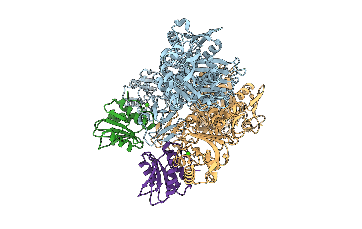
1NBW image
Deposition Date 2002-12-04
Release Date 2003-01-14
Last Version Date 2024-10-16
Entry Detail
PDB ID:
1NBW
Keywords:
Title:
Glycerol dehydratase reactivase
Biological Source:
Source Organism:
Host Organism:
Method Details:
Experimental Method:
Resolution:
2.40 Å
R-Value Free:
0.27
R-Value Work:
0.23
Space Group:
P 43 21 2
Macromolecular Entities
Structures with similar UniProt ID
Protein Blast
Polymer Type:polypeptide(L)
Molecule:GLYCEROL DEHYDRATASE REACTIVASE ALPHA SUBUNIT
Gene (Uniprot):dhaB4
Chain IDs:A, C
Chain Length:607
Number of Molecules:2
Biological Source:Klebsiella pneumoniae
Structures with similar UniProt ID
Protein Blast
Polymer Type:polypeptide(L)
Molecule:GLYCEROL DEHYDRATASE REACTIVASE BETA SUBUNIT
Gene (Uniprot):FXN67_07680
Chain IDs:B, D
Chain Length:117
Number of Molecules:2
Biological Source:Klebsiella pneumoniae
Ligand Molecules
Primary Citation
Structure of glycerol dehydratase reactivase: A new type of molecular chaperone
Structure 11 109 119 (2003)
PMID: 12517345 DOI: 10.1016/S0969-2126(02)00935-8

Abstact

The function of glycerol dehydratase (GDH) reactivase is to remove damaged coenzyme B(12) from GDH that has suffered mechanism-based inactivation. The structure of GDH reactivase from Klebsiella pneumoniae was determined at 2.4 A resolution by the single isomorphous replacement with anomalous signal (SIR/AS) method. Each tetramer contains two elongated 63 kDa alpha subunits and two globular 14 kDa beta subunits. The alpha subunit contains structural features resembling both GroEL and Hsp70 groups of chaperones, and it appears chaperone like in its interactions with ATP. The fold of the beta subunit resembles that of the beta subunit of glycerol dehydratase, except that it lacks some coenzyme B(12) binding elements. A hypothesis for the reactivation mechanism of reactivase is proposed based on these structural features.

Legend

Protein

Chemical

Disease

Primary Citation of related structures