Deposition Date
2002-12-03
Release Date
2003-02-18
Last Version Date
2024-02-14
Entry Detail
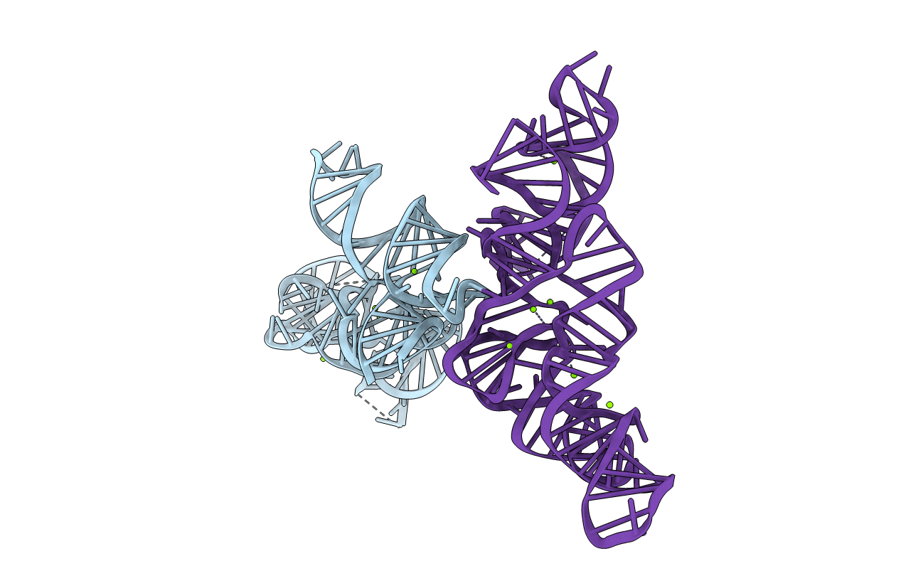
Biological Source:
Source Organism(s):
Bacillus subtilis (Taxon ID: 1423)
Expression System(s):
Method Details:
Experimental Method:
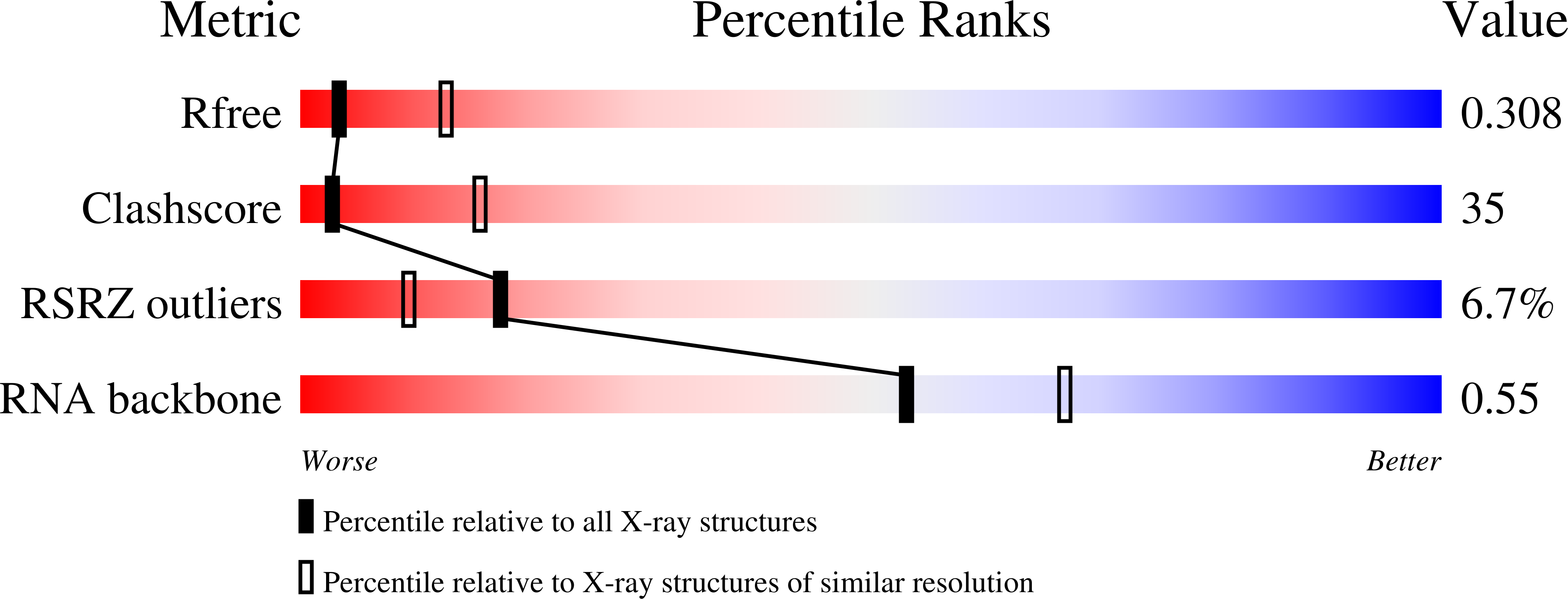
Resolution:
3.15 Å
R-Value Free:
0.30
R-Value Work:
0.28
R-Value Observed:
0.28
Space Group:
C 2 2 21