Deposition Date
2002-12-02
Release Date
2003-02-18
Last Version Date
2024-10-16
Entry Detail
PDB ID:
1NB5
Keywords:
Title:
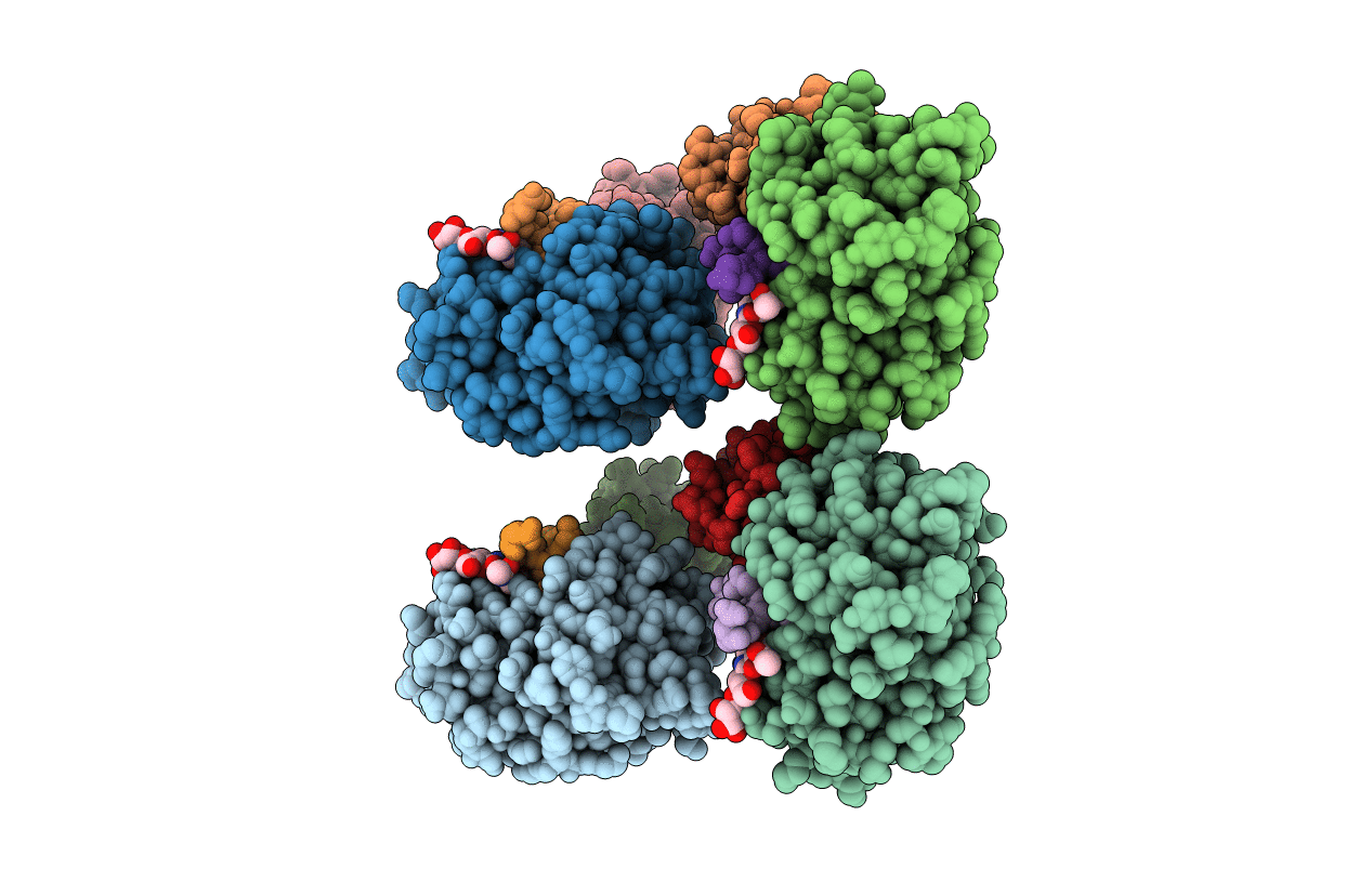
Crystal structure of stefin A in complex with cathepsin H
Biological Source:
Source Organism(s):
Homo sapiens (Taxon ID: 9606)
Sus scrofa (Taxon ID: 9823)
Sus scrofa (Taxon ID: 9823)
Expression System(s):
Method Details:
Experimental Method:
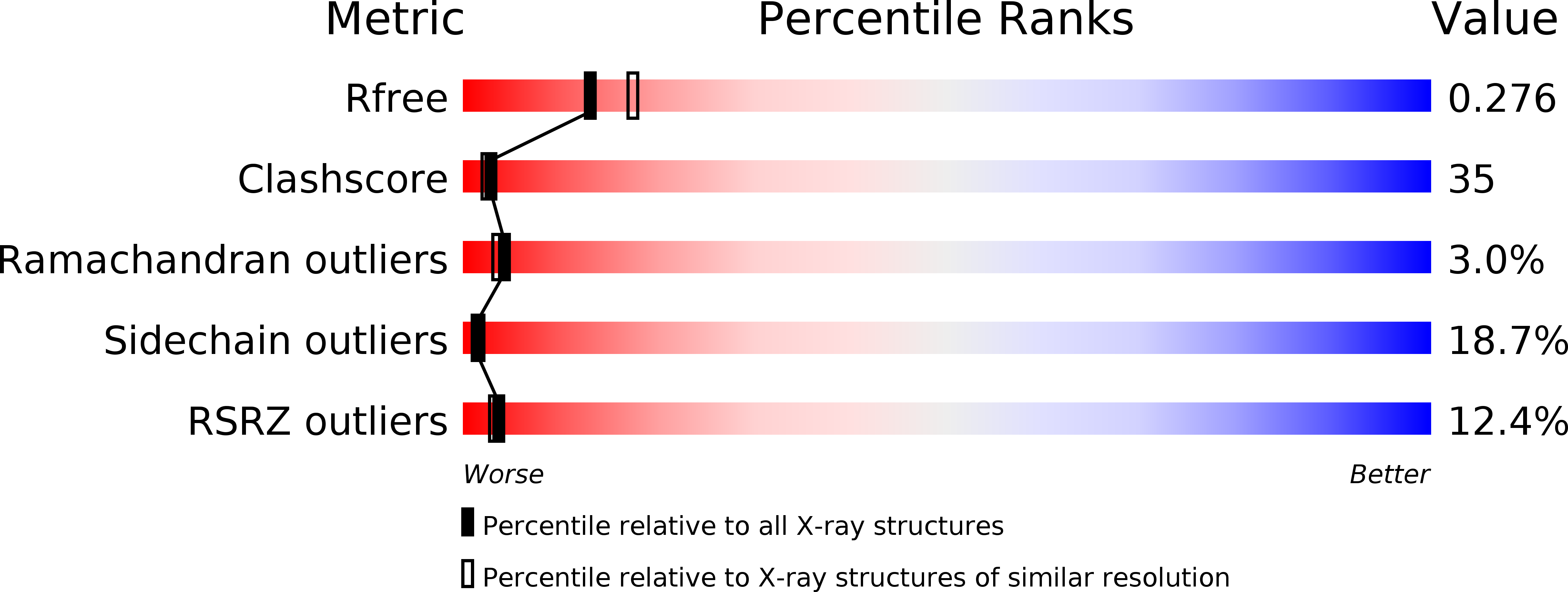
Resolution:
2.40 Å
R-Value Free:
0.27
R-Value Work:
0.23
Space Group:
P 21 21 21