
Deposition Date
1994-02-28
Release Date
1995-09-15
Last Version Date
2024-02-14
Entry Detail
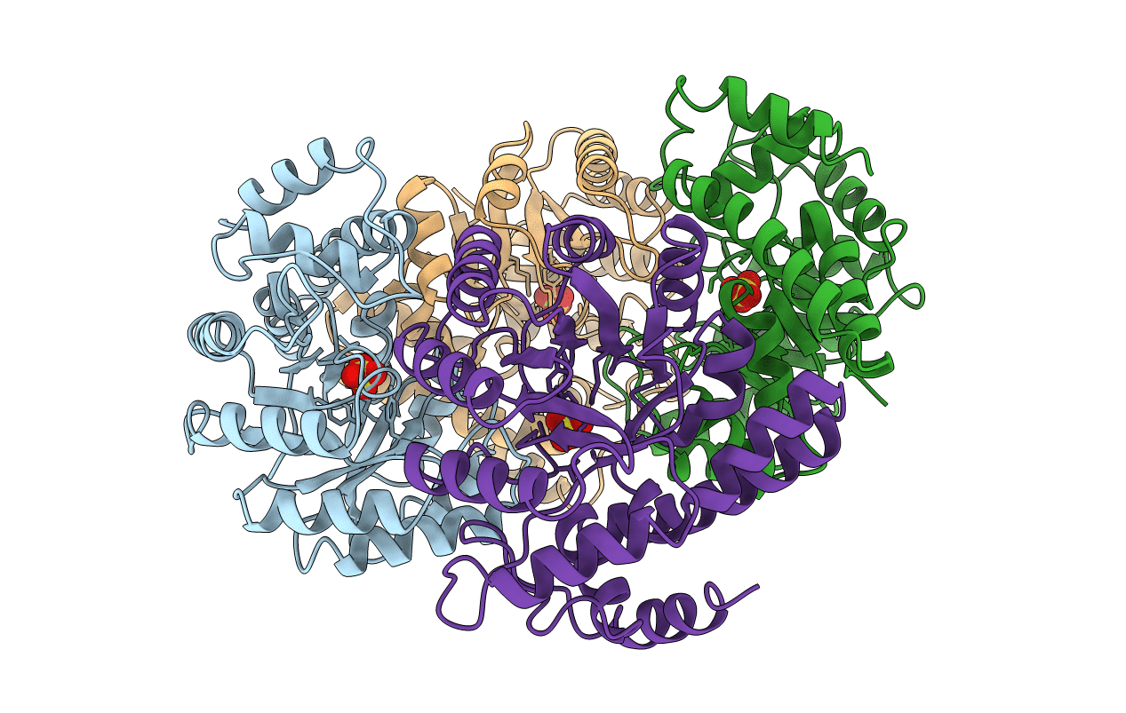
PDB ID:
1NAL
Keywords:
Title:
THE THREE-DIMENSIONAL STRUCTURE OF N-ACETYLNEURAMINATE LYASE FROM ESCHERICHIA COLI
Biological Source:
Source Organism(s):
Escherichia coli (Taxon ID: 562)
Method Details:
Experimental Method:
Resolution:
2.20 Å
R-Value Work:
0.20
R-Value Observed:
0.20
Space Group:
P 32 2 1
