1N9N image
Deposition Date 2002-11-25
Release Date 2003-06-17
Last Version Date 2023-10-25
Entry Detail
PDB ID:
1N9N
Title:
Crystal structure of the Phot-LOV1 domain from Chlamydomonas reinhardtii in illuminated state. Data set of a single crystal.
Biological Source:
Source Organism:
Host Organism:
Method Details:
Experimental Method:
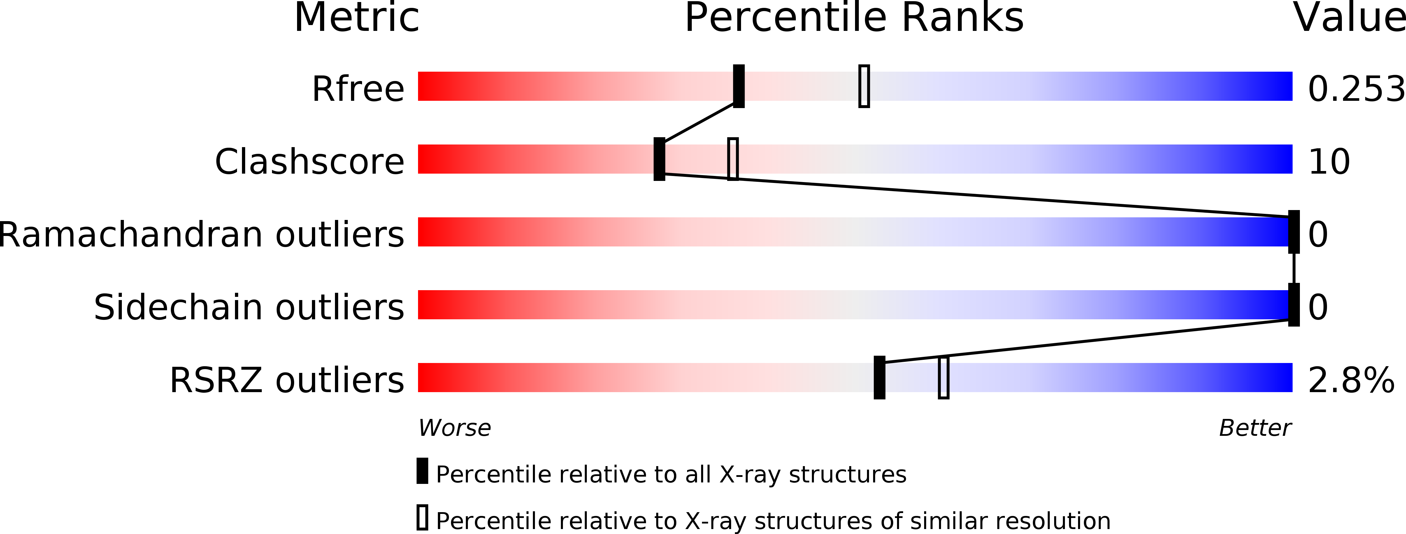
Resolution:
2.30 Å
R-Value Free:
0.26
R-Value Work:
0.21
R-Value Observed:
0.21
Space Group:
P 65 2 2
Macromolecular Entities
Structures with similar UniProt ID
Protein Blast
Polymer Type:polypeptide(L)
Molecule:putative blue light receptor
Chain IDs:A
Chain Length:109
Number of Molecules:1
Biological Source:Chlamydomonas reinhardtii
Ligand Molecules
Primary Citation
Crystal structures and molecular mechanism of a light-induced signaling switch: The Phot-LOV1 domain from Chlamydomonas reinhardtii.
Biophys.J. 84 2474 2482 (2003)
PMID: 12668455 DOI: 10.1016/S0006-3495(03)75052-8

Abstact

Phot proteins (phototropins and homologs) are blue-light photoreceptors that control mechanical processes like phototropism, chloroplast relocation, or guard-cell opening in plants. Phot receptors consist of two flavin mononucleotide (FMN)-binding light, oxygen, or voltage (LOV) domains and a C-terminal serine/threonine kinase domain. We determined crystal structures of the LOV1 domain of Phot1 from the green alga Chlamydomonas reinhardtii in the dark and illuminated state to 1.9 A and 2.8 A resolution, respectively. The structure resembles that of LOV2 from Adiantum (Crosson, S. and K. Moffat. 2001. PROC: Natl. Acad. Sci. USA. 98:2995-3000). In the resting dark state of LOV1, the reactive Cys-57 is present in two conformations. Blue-light absorption causes formation of a proposed active signaling state that is characterized by a covalent bond between the flavin C4a and the thiol of Cys-57. There are differences around the FMN chromophore but no large overall conformational changes. Quantum chemical calculations based on the crystal structures revealed the electronic distribution in the active site during the photocycle. The results suggest trajectories for electrons, protons, and the active site cysteine and offer an interpretation of the reaction mechanism.

Legend

Protein

Chemical

Disease

Primary Citation of related structures