Deposition Date
2002-11-21
Release Date
2003-03-04
Last Version Date
2024-02-14
Entry Detail
PDB ID:
1N8T
Keywords:
Title:
The crystal structure of phosphoglucose isomerase from rabbit muscle
Biological Source:
Source Organism(s):
Oryctolagus cuniculus (Taxon ID: 9986)
Method Details:
Experimental Method:
Resolution:
2.50 Å
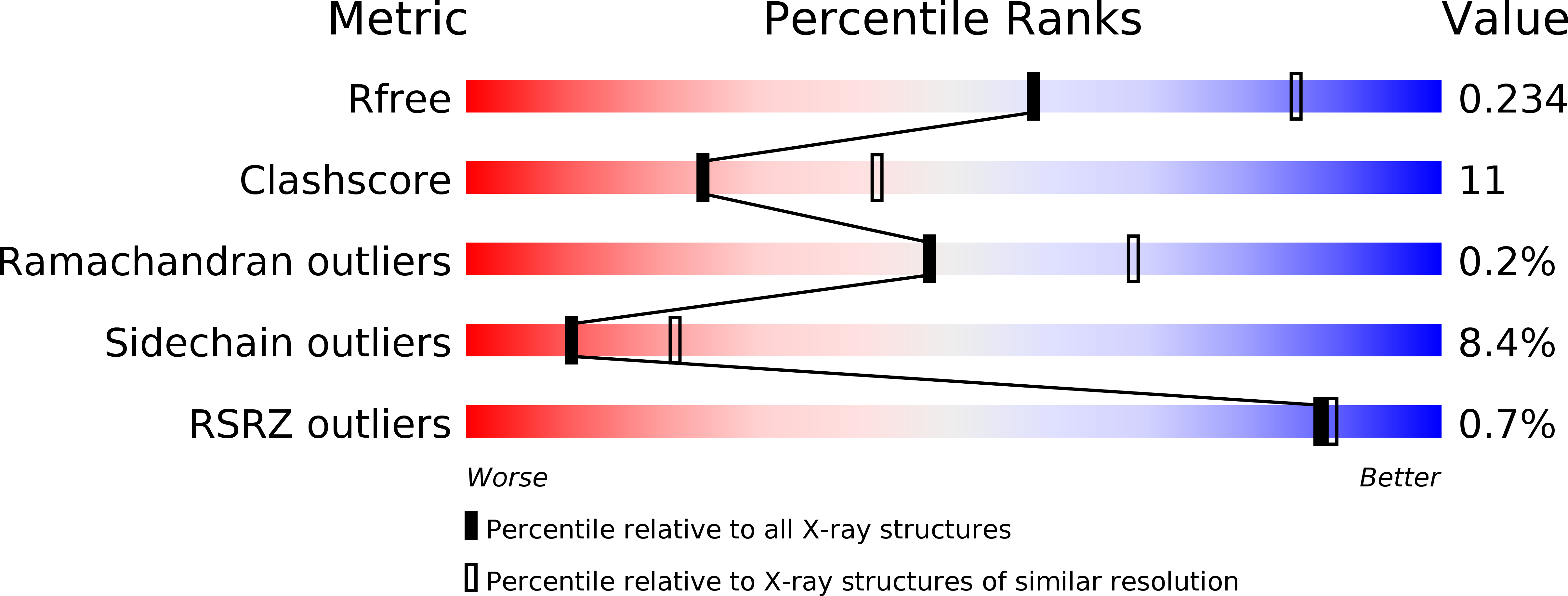
R-Value Free:
0.23
R-Value Work:
0.17
R-Value Observed:
0.18
Space Group:
C 2 2 21