Deposition Date
2002-11-18
Release Date
2003-04-08
Last Version Date
2024-02-14
Entry Detail
PDB ID:
1N7V
Keywords:
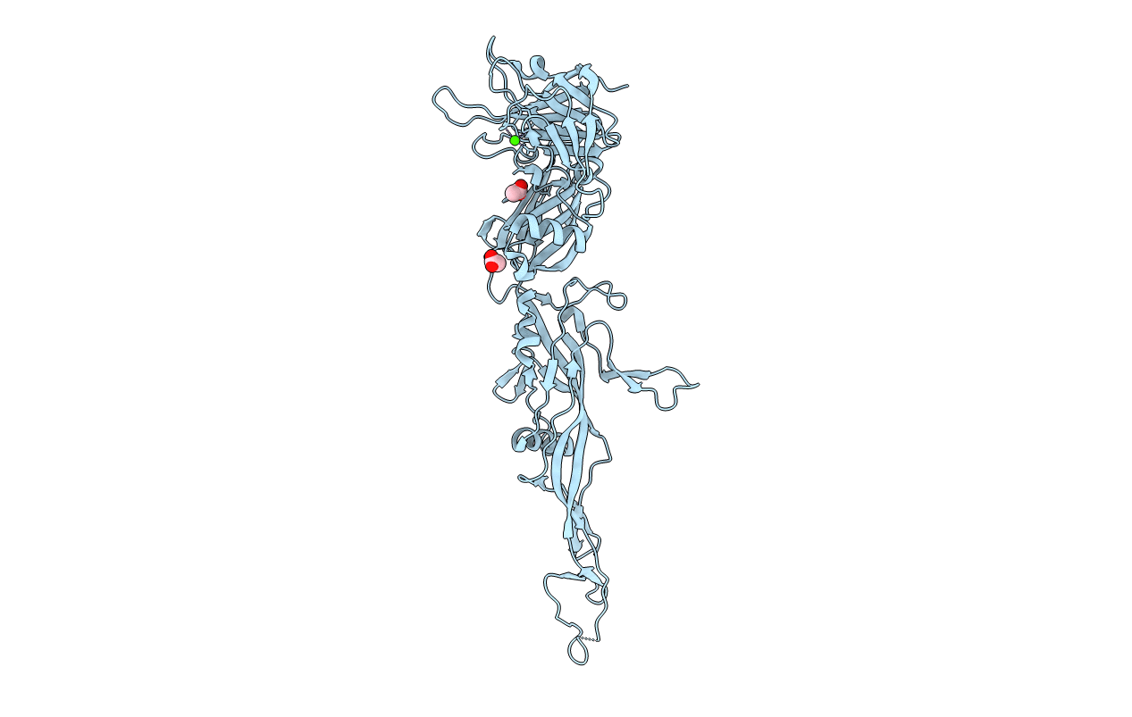
Title:
THE RECEPTOR-BINDING PROTEIN P2 OF BACTERIOPHAGE PRD1: CRYSTAL FORM III
Biological Source:
Source Organism(s):
Enterobacteria phage PRD1 (Taxon ID: 10658)
Expression System(s):
Method Details:
Experimental Method:
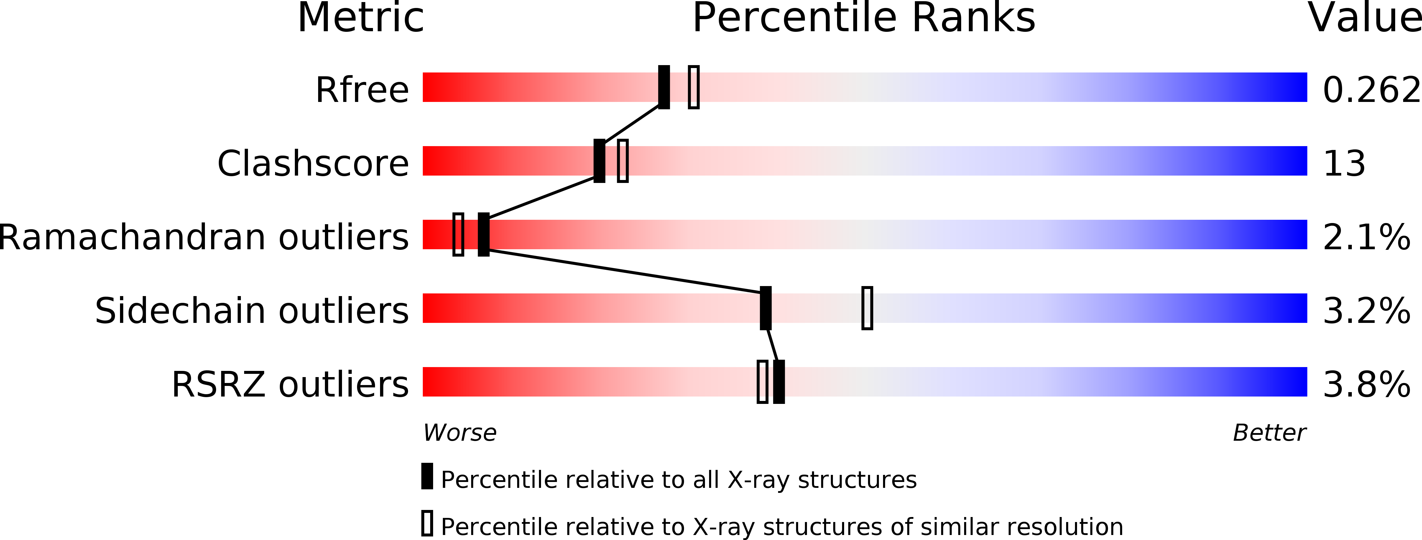
Resolution:
2.20 Å
R-Value Free:
0.26
R-Value Work:
0.22
R-Value Observed:
0.22
Space Group:
C 2 2 21