1N5Z image
Deposition Date 2002-11-08
Release Date 2002-12-11
Last Version Date 2024-03-13
Entry Detail
PDB ID:
1N5Z
Title:
Complex structure of Pex13p SH3 domain with a peptide of Pex14p
Biological Source:
Source Organism:
Saccharomyces cerevisiae (Taxon ID: 4932)
(Taxon ID: )
Method Details:
Experimental Method:
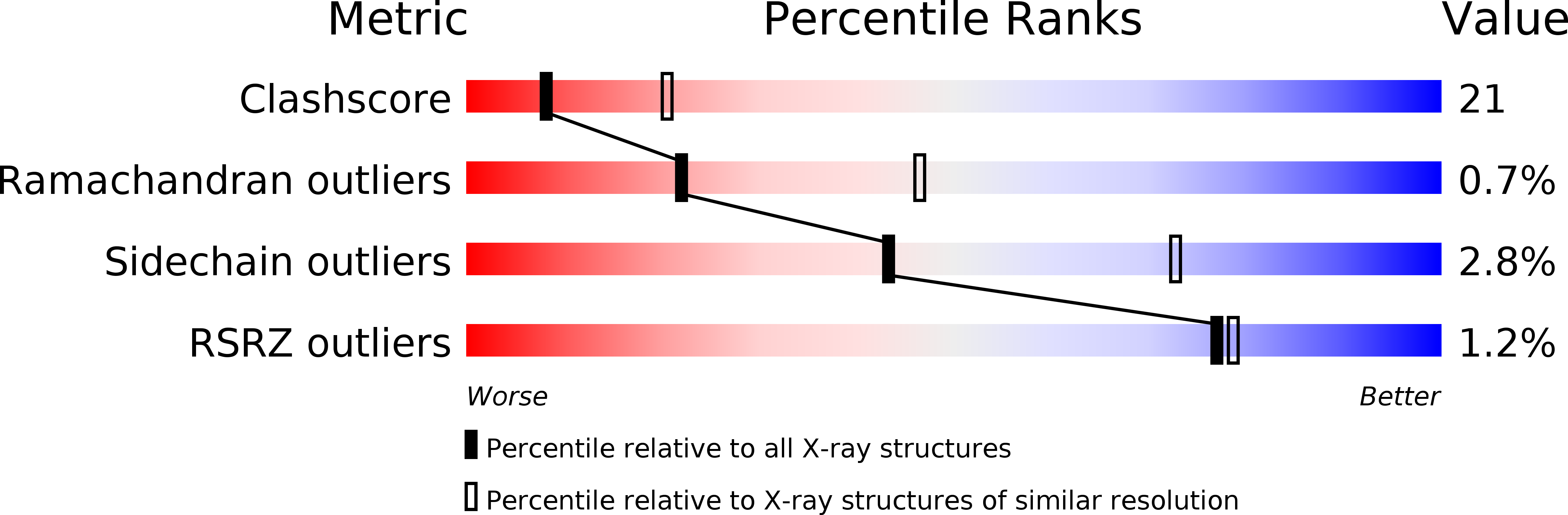
Resolution:
2.70 Å
R-Value Free:
0.27
R-Value Work:
0.24
R-Value Observed:
0.24
Space Group:
P 21 21 21
Macromolecular Entities
Polymer Type:polypeptide(L)
Molecule:Peroxisomal membrane protein PAS20
Gene (Uniprot):PEX13
Chain IDs:A, C (auth: B)
Chain Length:92
Number of Molecules:2
Biological Source:Saccharomyces cerevisiae
Polymer Type:polypeptide(L)
Molecule:14-mer peptide from Peroxisomal membrane protein PEX14
Gene (Uniprot):PEX14
Chain IDs:B (auth: P), D (auth: Q)
Chain Length:14
Number of Molecules:2
Biological Source:
Primary Citation
Topography for Independent Binding of alpha-Helical and PPII-Helical Ligands to a Peroxisomal SH3 Domain
MOL.CELL 10 1007 1017 (2002)
PMID: 12453410 DOI: 10.1016/S1097-2765(02)00749-9

Abstact

While the function of most small signaling domains is confined to binary ligand interactions, the peroxisomal Pex13p SH3 domain has the unique capacity of binding to two different ligands, Pex5p and Pex14p. We have used this domain as a model to decipher its structurally independent ligand binding sites. By the combined use of X-ray crystallography, NMR spectroscopy, and circular dichroism, we show that the two ligands bind in unrelated conformations to patches located at opposite surfaces of this SH3 domain. Mutations in the Pex13p SH3 domain that abolish interactions within the Pex13p-Pex5p interface specifically impair PTS1-dependent protein import into yeast peroxisomes.

Legend

Protein

Chemical

Disease

Primary Citation of related structures