
Deposition Date
2002-11-04
Release Date
2003-01-21
Last Version Date
2024-02-14
Entry Detail
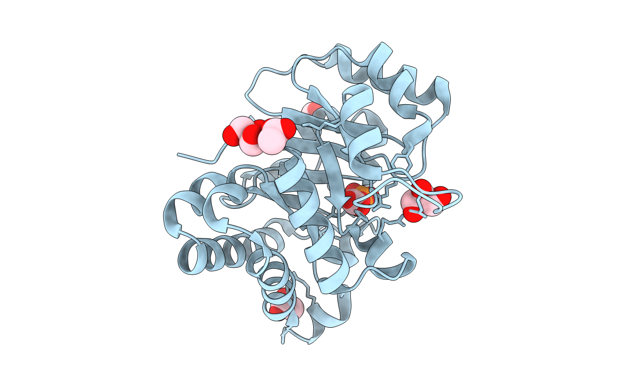
PDB ID:
1N55
Keywords:
Title:
0.83A resolution structure of the E65Q mutant of Leishmania mexicana triosephosphate isomerase complexed with 2-phosphoglycolate
Biological Source:
Source Organism(s):
Leishmania mexicana (Taxon ID: 5665)
Expression System(s):
Method Details:
Experimental Method:
Resolution:
0.83 Å
R-Value Free:
0.10
R-Value Work:
0.09
R-Value Observed:
0.09
Space Group:
C 1 2 1
