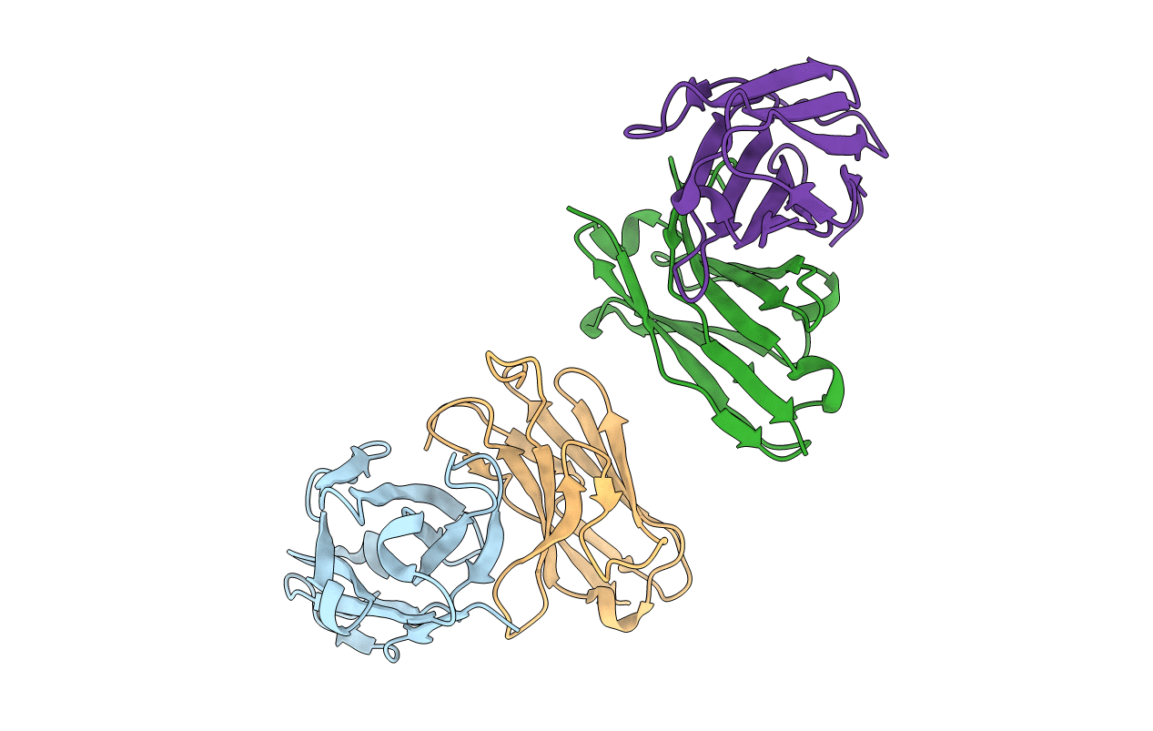
1N4X image
Deposition Date 2002-11-02
Release Date 2003-06-10
Last Version Date 2024-11-13
Entry Detail
PDB ID:
1N4X
Keywords:
Title:
Structure of scFv 1696 at acidic pH
Biological Source:
Source Organism:
Mus musculus (Taxon ID: 10090)
Host Organism:
Method Details:
Experimental Method:
Resolution:
1.70 Å
R-Value Free:
0.25
R-Value Work:
0.22
R-Value Observed:
0.22
Space Group:
P 21 21 21
Macromolecular Entities
Polymer Type:polypeptide(L)
Molecule:immunoglobulin heavy chain variable region
Chain IDs:B (auth: H), D (auth: I)
Chain Length:120
Number of Molecules:2
Biological Source:Mus musculus
Polymer Type:polypeptide(L)
Molecule:immunoglobulin kappa chain variable region
Chain IDs:A (auth: L), C (auth: M)
Chain Length:113
Number of Molecules:2
Biological Source:Mus musculus
Ligand Molecules
Primary Citation
Structure of a single-chain Fv fragment of an antibody that inhibits the HIV-1 and HIV-2 proteases.
Acta Crystallogr.,Sect.D 59 955 957 (2003)
PMID: 12777823 DOI: 10.1107/S0907444903003597

Abstact

The monoclonal antibody 1696, which was raised against the HIV-1 protease, inhibits the catalytic activity of the enzyme from both the HIV-1 and HIV-2 strains. The antibody cross-reacts with peptides containing the N-terminus of the enzyme, which is highly conserved between these strains. The crystal structure of a single-chain Fv fragment of 1696 (scFv-1696) in the non-complexed form, solved at 1.7 A resolution, is compared with the previously reported non-complexed Fab-1696 and antigen-bound scFv-1696 structures. Large conformational changes in the third hypervariable region of the heavy chain and differences in relative orientation of the variable domains are observed between the different structures.

Legend

Protein

Chemical

Disease

Primary Citation of related structures