Deposition Date
2002-10-31
Release Date
2003-01-07
Last Version Date
2024-02-14
Entry Detail
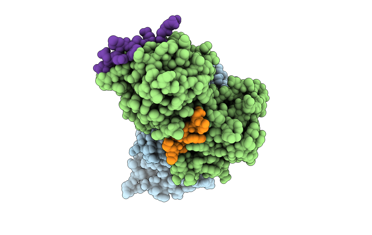
PDB ID:
1N4M
Keywords:
Title:
Structure of Rb tumor suppressor bound to the transactivation domain of E2F-2
Biological Source:
Source Organism(s):
Homo sapiens (Taxon ID: 9606)
Expression System(s):
Method Details:
Experimental Method:
Resolution:
2.20 Å
R-Value Free:
0.28
R-Value Work:
0.22
R-Value Observed:
0.22
Space Group:
P 1