Deposition Date
2002-10-30
Release Date
2002-11-13
Last Version Date
2024-02-14
Entry Detail
PDB ID:
1N45
Keywords:
Title:
X-RAY CRYSTAL STRUCTURE OF HUMAN HEME OXYGENASE-1 (HO-1) IN COMPLEX WITH ITS SUBSTRATE HEME
Biological Source:
Source Organism(s):
Homo sapiens (Taxon ID: 9606)
Expression System(s):
Method Details:
Experimental Method:
Resolution:
1.50 Å
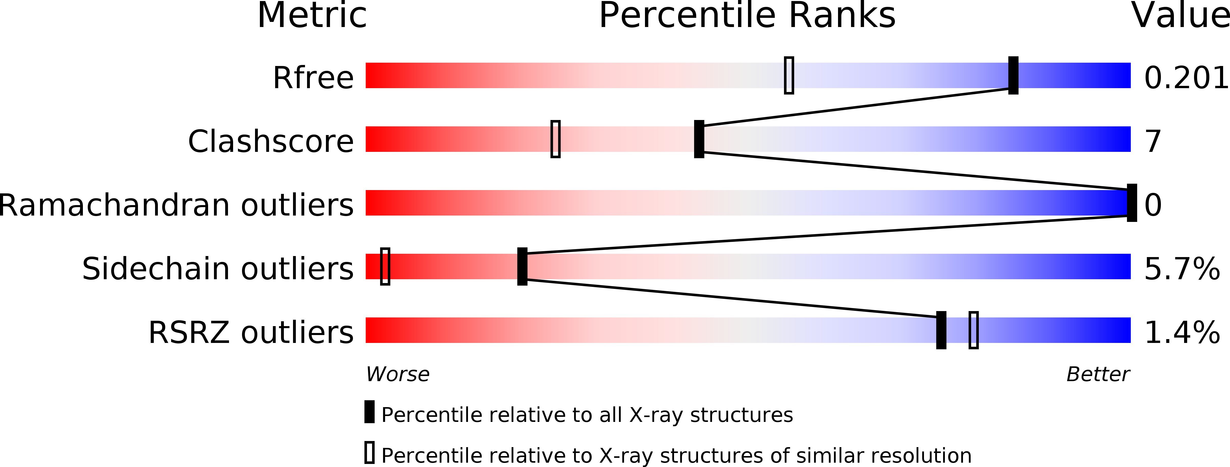
R-Value Free:
0.21
R-Value Work:
0.15
Space Group:
P 1 21 1