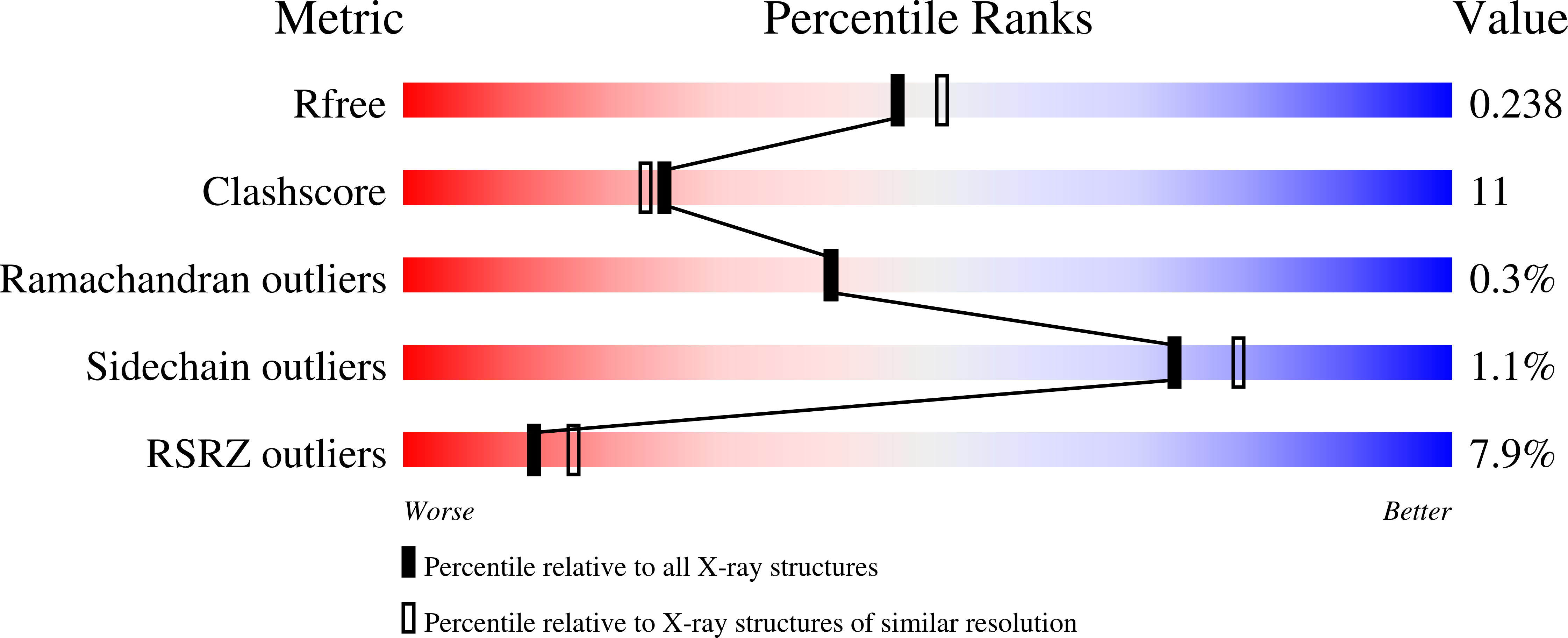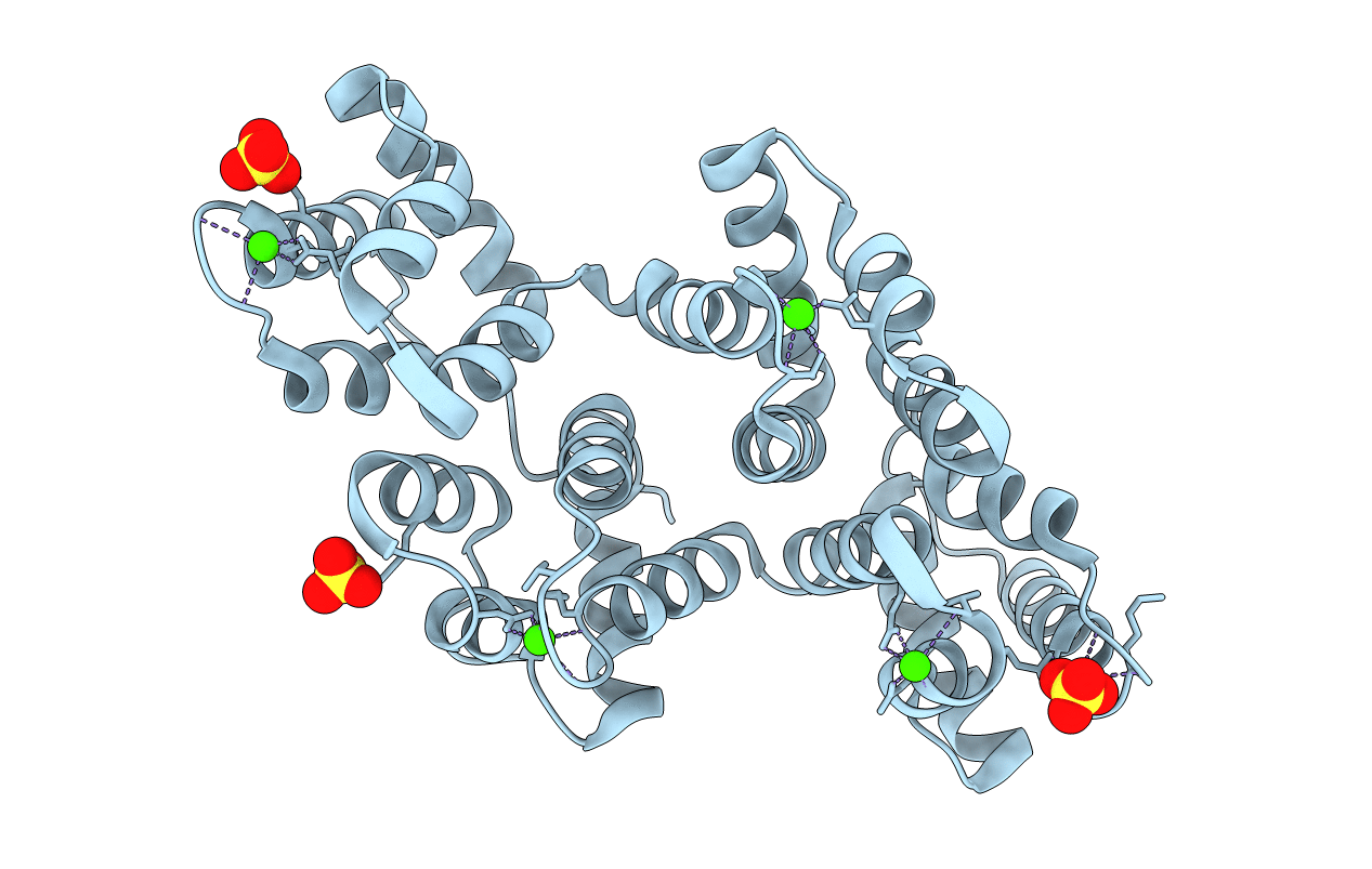
1N42 image
Deposition Date 2002-10-30
Release Date 2003-02-04
Last Version Date 2024-02-14
Entry Detail
PDB ID:
1N42
Title:
Crystal Structure of Annexin V R149E Mutant
Biological Source:
Source Organism:
Host Organism:
Method Details:
Experimental Method:
Resolution:
2.10 Å
R-Value Free:
0.23
R-Value Work:
0.19
R-Value Observed:
0.19
Space Group:
H 3
Macromolecular Entities
Structures with similar UniProt ID
Protein Blast
Polymer Type:polypeptide(L)
Molecule:Annexin V
Gene (Uniprot):Anxa5
Mutagens:R149E
Chain IDs:A
Chain Length:319
Number of Molecules:1
Biological Source:Rattus norvegicus
Primary Citation
Interfacial basic cluster in annexin V couples phospholipid binding and trimer formation on membrane surfaces
J.Biol.Chem. 278 2437 2443 (2003)
PMID: 12401794 DOI: 10.1074/jbc.M210286200

Abstact

Annexin V is an abundant eukaryotic protein that binds phospholipid membranes in a Ca(2+)-dependent manner. In the present studies, site-directed mutagenesis was combined with x-ray crystallography and solution liposome binding assays to probe the functional role of a cluster of interfacial basic residues in annexin V. Four mutants were investigated: R23E, K27E, R61E, and R149E. All four mutants exhibited a significant reduction in adsorption to phospholipid membranes relative to the wild-type protein, and the R23E mutation was the most deleterious. Crystal structures of wild-type and mutant proteins were similar except for local changes in salt bridges involving basic cluster residues. The combined data indicate that Arg(23) is a major determinant for interfacial phospholipid binding and participates in an intermolecular salt bridge that is key for trimer formation on the membrane surface. Together, crystallographic and solution data provide evidence that the interfacial basic cluster is a locus where trimerization is synergistically coupled to membrane phospholipid binding.

Legend

Protein

Chemical

Disease

Primary Citation of related structures