Deposition Date
2002-10-24
Release Date
2003-04-08
Last Version Date
2024-02-14
Entry Detail
PDB ID:
1N2V
Keywords:
Title:
Crystal Structure of TGT in complex with 2-Butyl-5,6-dihydro-1H-imidazo[4,5-d]pyridazine-4,7-dione
Biological Source:
Source Organism(s):
Zymomonas mobilis (Taxon ID: 542)
Expression System(s):
Method Details:
Experimental Method:
Resolution:
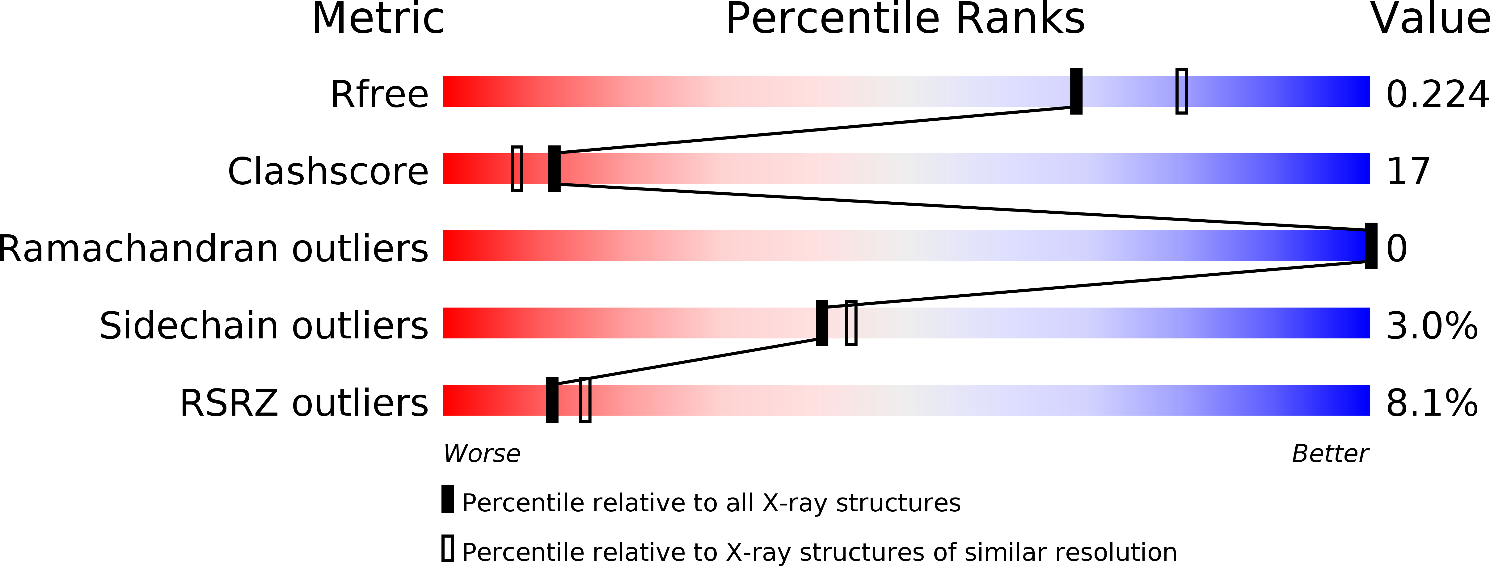
2.10 Å
R-Value Free:
0.23
R-Value Work:
0.18
Space Group:
C 1 2 1