1N1H image
Deposition Date 2002-10-17
Release Date 2002-12-25
Last Version Date 2024-04-03
Entry Detail
PDB ID:
1N1H
Keywords:
Title:
Initiation complex of polymerase lambda3 from reovirus
Biological Source:
Source Organism:
Mammalian orthoreovirus 3 (Taxon ID: 10886)
(Taxon ID: )
Host Organism:
Method Details:
Experimental Method:
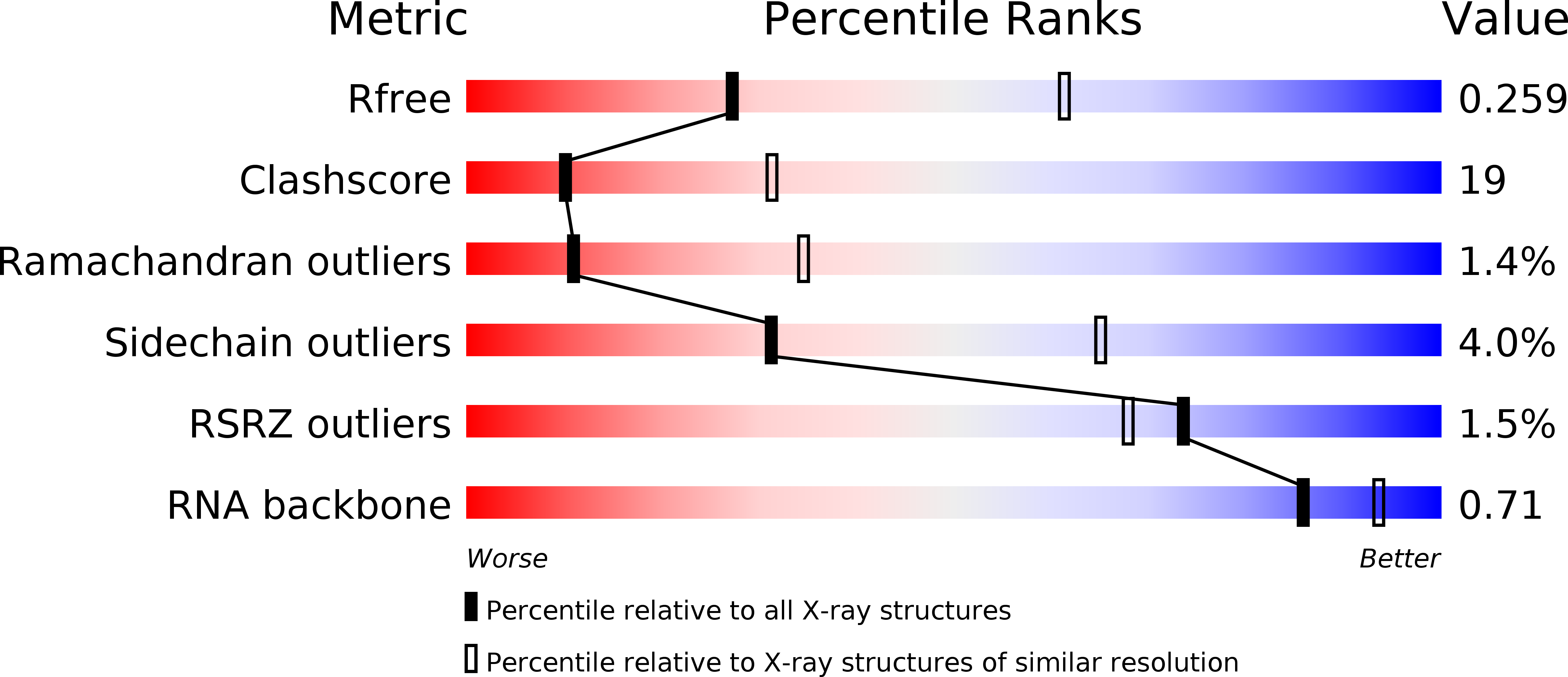
Resolution:
2.80 Å
R-Value Free:
0.26
R-Value Work:
0.21
R-Value Observed:
0.21
Space Group:
P 21 21 21
Macromolecular Entities
Polymer Type:polypeptide(L)
Molecule:Minor core protein lambda 3
Chain IDs:B (auth: A)
Chain Length:1267
Number of Molecules:1
Biological Source:Mammalian orthoreovirus 3
Polymer Type:polyribonucleotide
Molecule:5'-R(*AP*UP*UP*AP*GP*C)-3'
Chain IDs:A (auth: B)
Chain Length:6
Number of Molecules:1
Biological Source:
Primary Citation
RNA Synthesis in a Cage--Structural Studies of Reovirus Polymerase [lambda] 3
Cell(Cambridge,Mass.) 111 733 745 (2002)
PMID: 12464184 DOI: 10.1016/S0092-8674(02)01110-8

Abstact

The reovirus polymerase and those of other dsRNA viruses function within the confines of a protein capsid to transcribe the tightly packed dsRNA genome segments. The crystal structure of the reovirus polymerase, lambda3, determined at 2.5 A resolution, shows a fingers-palm-thumb core, similar to those of other viral polymerases, surrounded by major N- and C-terminal elaborations, which create a cage-like structure, with four channels leading to the catalytic site. This "caged" polymerase has allowed us to visualize the results of several rounds of RNA polymerization directly in the crystals. A 5' cap binding site on the surface of lambda3 suggests a template retention mechanism by which attachment of the 5' end of the plus-sense strand facilitates insertion of the 3' end of the minus-sense strand into the template channel.

Legend

Protein

Chemical

Disease

Primary Citation of related structures