1N11 image
Deposition Date 2002-10-16
Release Date 2002-12-11
Last Version Date 2024-02-14
Entry Detail
PDB ID:
1N11
Title:
D34 REGION OF HUMAN ANKYRIN-R AND LINKER
Biological Source:
Source Organism:
Homo sapiens (Taxon ID: 9606)
Host Organism:
Method Details:
Experimental Method:
Resolution:
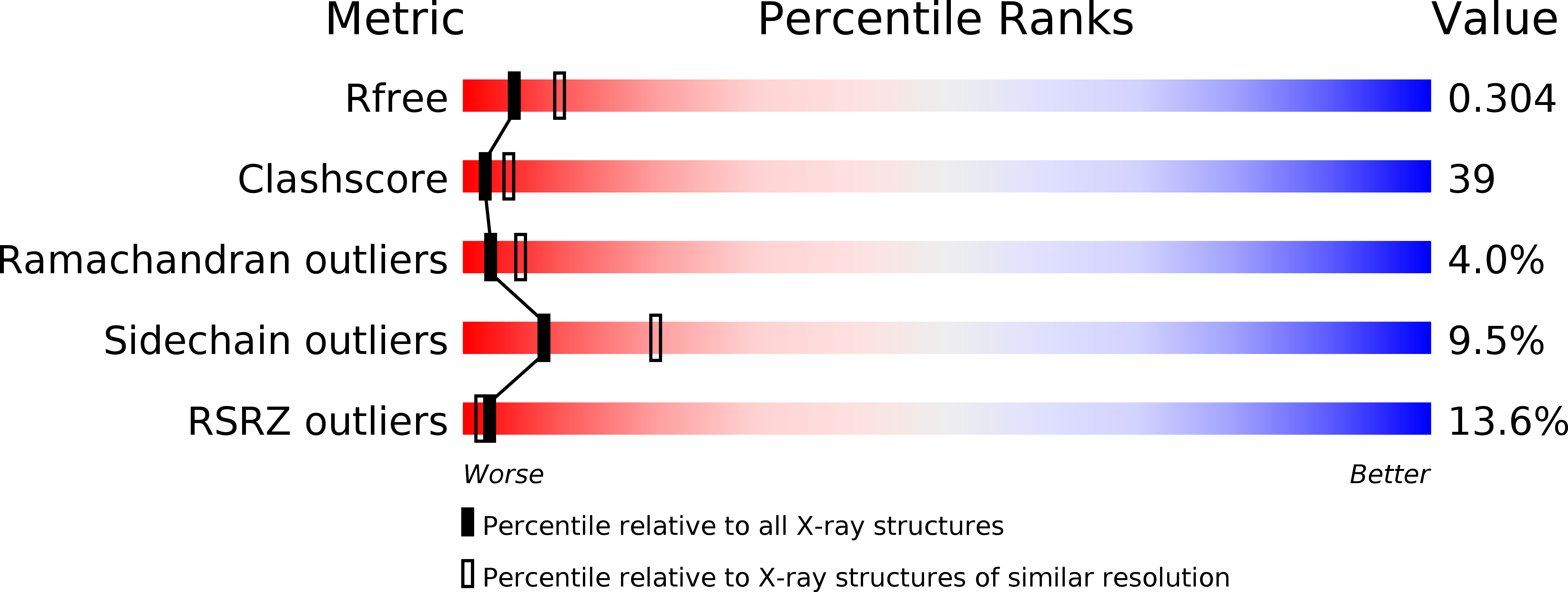
2.70 Å
R-Value Free:
0.30
R-Value Work:
0.31
R-Value Observed:
0.31
Space Group:
P 63 2 2
Macromolecular Entities
Polymer Type:polypeptide(L)
Molecule:Ankyrin
Gene (Uniprot):ANK1
Chain IDs:A
Chain Length:437
Number of Molecules:1
Biological Source:Homo sapiens
Primary Citation
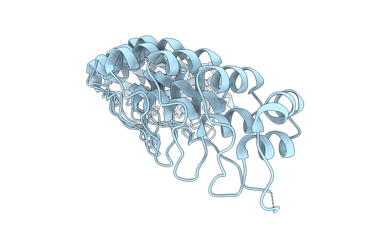
Crystal structure of a 12 ANK repeat stack from human ankyrinR
Embo J. 21 6387 6396 (2002)
PMID: 12456646 DOI: 10.1093/emboj/cdf651

Abstact

Ankyrins are multifunctional adaptors that link specific proteins to the membrane-associated, spectrin- actin cytoskeleton. The N-terminal, 'membrane-binding' domain of ankyrins contains 24 ANK repeats and mediates most binding activities. Repeats 13-24 are especially active, with known sites of interaction for the Na/K ATPase, Cl/HCO(3) anion exchanger, voltage-gated sodium channel, clathrin heavy chain and L1 family cell adhesion molecules. Here we report the crystal structure of a human ankyrinR construct containing ANK repeats 13-24 and a portion of the spectrin-binding domain. The ANK repeats are observed to form a contiguous spiral stack with which the spectrin-binding domain fragment associates as an extended strand. The structural information has been used to construct models of all 24 repeats of the membrane-binding domain as well as the interactions of the repeats with the Cl/HCO(3) anion exchanger and clathrin. These models, together with available binding studies, suggest that ion transporters such as the anion exchanger associate in a large central cavity formed by the ANK repeat spiral, while clathrin and cell adhesion molecules associate with specific regions outside this cavity.

Legend

Protein

Chemical

Disease

Primary Citation of related structures