
Deposition Date
2002-10-07
Release Date
2003-01-28
Last Version Date
2024-11-20
Entry Detail
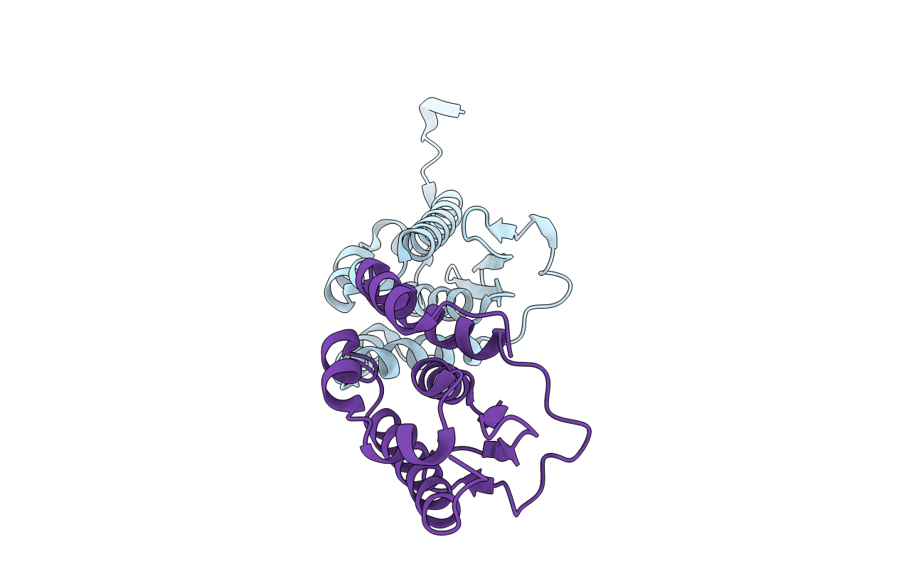
PDB ID:
1MZG
Keywords:
Title:
X-Ray Structure of SufE from E.coli Northeast Structural Genomics (NESG) Consortium Target ER30
Biological Source:
Source Organism(s):
Escherichia coli (Taxon ID: 562)
Expression System(s):
Method Details:
Experimental Method:
Resolution:
2.00 Å
R-Value Free:
0.25
R-Value Work:
0.20
Space Group:
P 21 21 21
