Deposition Date
1999-04-22
Release Date
1999-07-06
Last Version Date
2023-12-27
Entry Detail
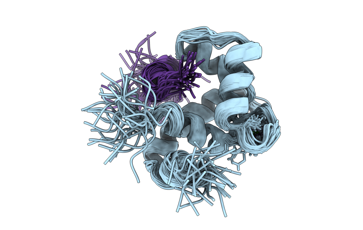
PDB ID:
1MXL
Keywords:
Title:
STRUCTURE OF CARDIAC TROPONIN C-TROPONIN I COMPLEX
Biological Source:
Source Organism(s):
Homo sapiens (Taxon ID: 9606)
Expression System(s):
Method Details:
Experimental Method:
Conformers Calculated:
100
Conformers Submitted:
40
Selection Criteria:
LEAST RESTRAINT VIOLATION