1MWN image
Entry Detail
PDB ID:
1MWN
Title:
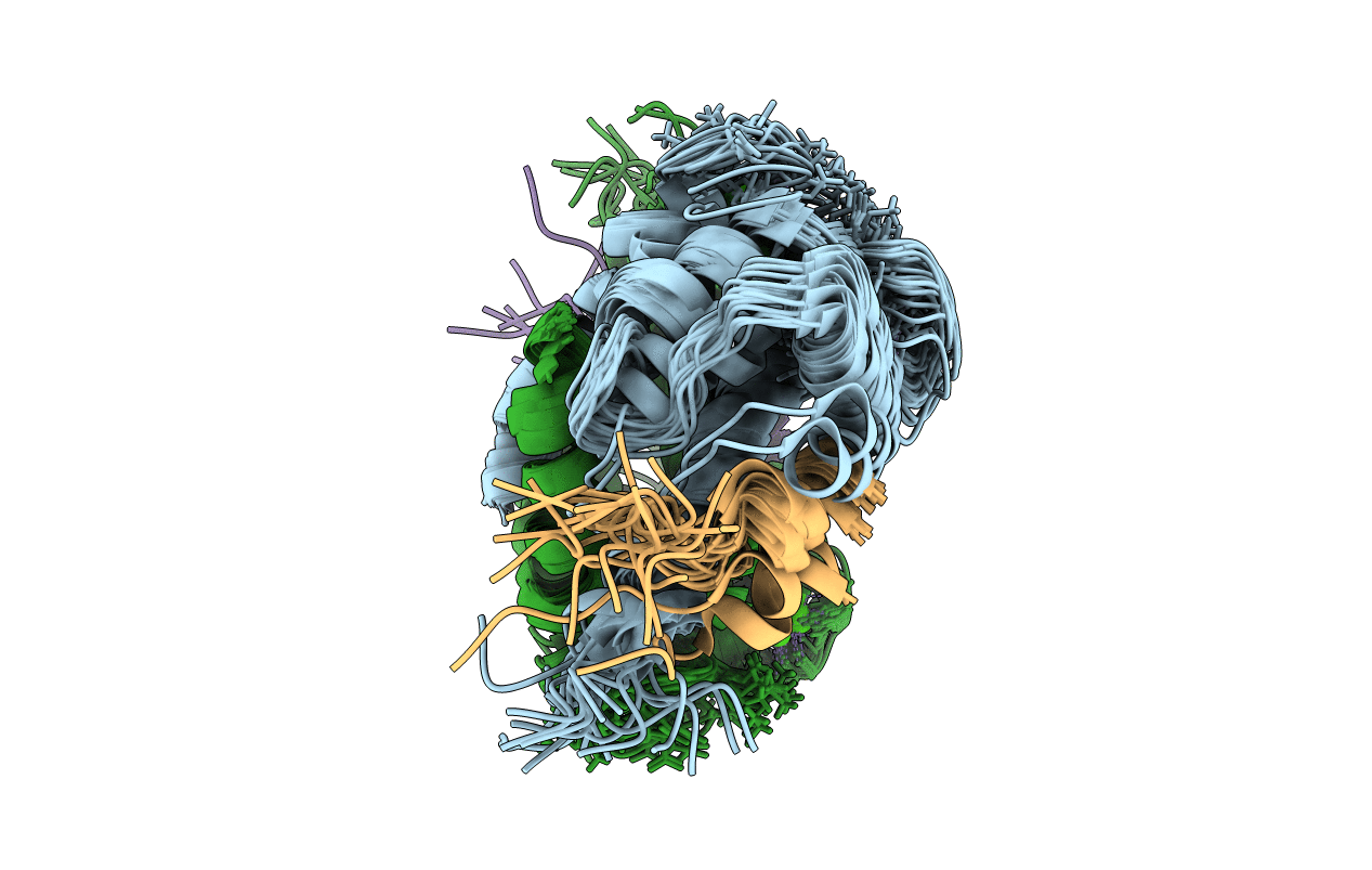
Solution NMR structure of S100B bound to the high-affinity target peptide TRTK-12
Biological Source:
Source Organism:
Host Organism:
PDB Version:
Deposition Date:
2002-09-30
Release Date:
2002-12-18
Method Details:
Experimental Method:
Conformers Calculated:
200
Conformers Submitted:
20
Selection Criteria:
structures with the lowest energy
Macromolecular Entities
Polymer Type:polypeptide(L)
Description:S-100 protein, beta chain
Chain IDs:A, B
Chain Length:92
Number of Molecules:2
Biological Source:Rattus norvegicus
Polymer Type:polypeptide(L)
Description:F-actin capping protein alpha-1 subunit
Chain IDs:C (auth: X), D (auth: Y)
Chain Length:12
Number of Molecules:2
Biological Source:
Ligand Molecules
Primary Citation

Abstact

The solution NMR structure is reported for Ca(2+)-loaded S100B bound to a 12-residue peptide, TRTK-12, from the actin capping protein CapZ (alpha1 or alpha2 subunit, residues 265-276: TRTKIDWNKILS). This peptide was discovered by Dimlich and co-workers by screening a bacteriophage random peptide display library, and it matches exactly the consensus S100B binding sequence ((K/R)(L/I)XWXXIL). As with other S100B target proteins, a calcium-dependent conformational change in S100B is required for TRTK-12 binding. The TRTK-12 peptide is an amphipathic helix (residues W7 to S12) in the S100B-TRTK complex, and helix 4 of S100B is extended by three or four residues upon peptide binding. However, helical TRTK-12 in the S100B-peptide complex is uniquely oriented when compared to the three-dimensional structures of other S100-peptide complexes. The three-dimensional structure of the S100B-TRTK peptide complex illustrates that residues in the S100B binding consensus sequence (K4, I5, W7, I10, L11) are all involved in the S100B-peptide interface, which can explain its orientation in the S100B binding pocket and its relatively high binding affinity. A comparison of the S100B-TRTK peptide structure to the structures of apo- and Ca(2+)-bound S100B illustrates that the binding site of TRTK-12 is buried in apo-S100B, but is exposed in Ca(2+)-bound S100B as necessary to bind the TRTK-12 peptide.

Legend

Protein

Chemical

Disease

Primary Citation of related structures