Deposition Date
2002-09-25
Release Date
2003-07-15
Last Version Date
2024-11-20
Entry Detail
PDB ID:
1MVE
Keywords:
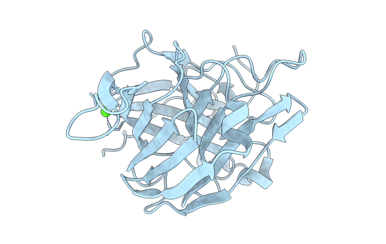
Title:
Crystal structure of a natural circularly-permutated jellyroll protein: 1,3-1,4-beta-D-glucanase from Fibrobacter succinogenes
Biological Source:
Source Organism(s):
Fibrobacter succinogenes (Taxon ID: 833)
Expression System(s):
Method Details:
Experimental Method:
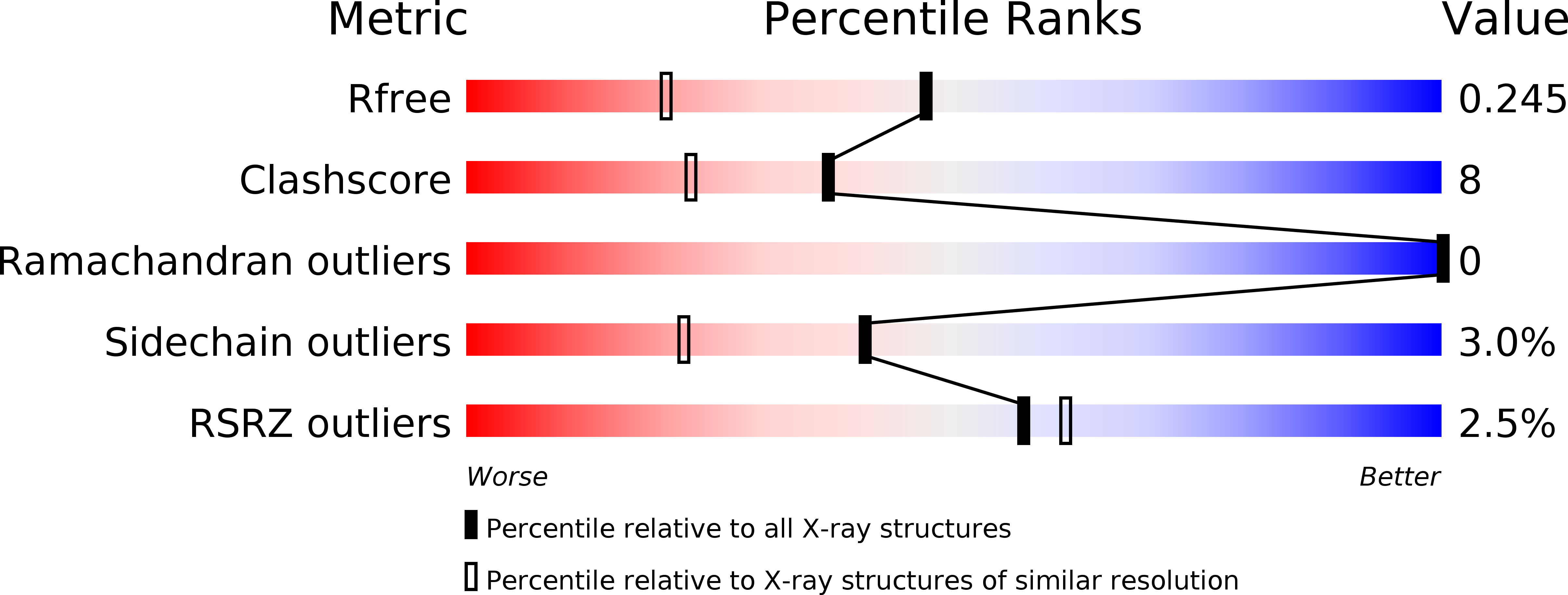
Resolution:
1.70 Å
R-Value Free:
0.23
R-Value Work:
0.19
R-Value Observed:
0.21
Space Group:
P 21 21 21